
Page 407 - The Veterinary Laboratory and Field Manual 3rd Edition
P. 407
376 Susan C. Cork
Musculoskeletal system four or five digits. It is important to examine
(see Figures 8.10, 8.11 and 8.12) the limbs carefully in an animal which has had a
history of lameness but if no obvious abnormali-
The musculature should be examined for evi- ties are present in the muscles, bone, joints or
dence of swelling or bruising. Note that bruises associated tendons/ligaments the problem may
can only occur ante-mortem but that autolytic have been in the nervous system. It is not easy
changes may occur after death. The musculo- to diagnose peripheral neurological damage at
skeletal system has the same basic structure in
all mammalian species but in birds the bones are necropsy without histological examination of
filled with air spaces which may connect with nerves, this is quite specialized and requires
internal air sacs. In some cases, broken bones specialist knowledge.
may result in entrance of infectious organisms
into the respiratory system of the bird and result Endocrine system
in septicaemia and death. Figure 8.11 illustrates
the general skeletal structure of the bovine. The endocrine system is a system of glands
Ruminants and horses have a fixed spinal col- which secretes hormones to regulate body func-
umn and strong neck ligaments. The lower limbs tions. The endocrine and neurological systems
of ruminant species and pigs have two main work together to maintain the body in its normal
digits as compared with a single main digit in physiological state. The control centre for most
the horse (Figure 8.12). Note that most gen- endocrine functions is the hypothalamus, which
eral lameness in the horse is usually in the foot. is located at the base of the brain. The hypo-
Examine the feet and lower limbs of a live horse thalamus controls the production and secretion
to identify the main structures illustrated. The of hormones in the pituitary gland which in turn
lower limbs of non-hoofed animals tend to have regulates other endocrine glands in a feedback
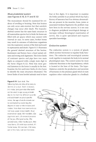
Figure 8.10 Goat skull. The
following features should be easy to
identify on a skull. Teeth: (i) incisor,
(m) molar, (pm) premolar (the teeth
can be used to age young animals
due to the fact that different juvenile
and adult teeth erupt at specific
times, the age of adult animals can
be estimated by examining the
degree of wear on the incisors and
molars. Note that most ruminants do
not have upper incisor teeth). (Exa)
external ear canal, (M) mandible, (Za)
zygomatic arch, (J) coronoid process
of the jaw, (Pm) premaxilla, (Pb)
parietal bone, (Ma) maxilla (jaw), (Nb)
nasal bone, (O) orbit (eye socket), (F)
frontal bone, (Cr) cranium, (Hb) bony
part of the horn, (Lb) lacrimal bone.
Vet Lab.indb 376 26/03/2019 10:26