
Page 406 - The Veterinary Laboratory and Field Manual 3rd Edition
P. 406
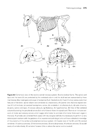
Pathology/cytology  375


                                                             B


                             A1                        A2


                                         C


                Figure 8.9  Schematic view of the bovine central nervous system. Bovine skeletal frame. The spinal cord
                (spc) and the brain (A) are protected by the vertebral column and the skull and are surrounded by three
                membranes (the meninges) and a layer of protective fluid. Illustrations (A, B and C) show some anatomical
                features of the brain, spinal column and vertebrae (v), respectively. (A) Lateral view (A2) and sagittal sec-
                tion (A1) of the brain, (a) cerebral hemisphere, cortex, (b) cerebellum, (c) olfactory bulb, (d) optic chiasma,
                (e) pons, spinal cord (spc), (f) corpus callosum, (g) thalamus, (h) hypothalamus. (B) View of the vertebral
                column illustrating the peripheral nerves (bn) which branch from the spinal cord. There is an intricate net-
                work of motor and sensory nerves which supply information to and take information from each organ in
                the body. If samples are collected from cases with neurological deficits it is necessary to perform a very
                detailed post-mortem with the guidance of an experienced pathologist who will have a detailed knowledge
                of the anatomy of the central and peripheral nervous system. (C) Caudal view of the 6th and 7th cervical
                vertebrae. (1) spinous process, (2) transverse process, (3) articular process, (4) articular process, (5) caudal
                cavity of vertebral body, (6) vertebral foramen (this is where the spinal cord is located).


       Vet Lab.indb   375                                                                  26/03/2019   10:26