
Page 416 - The Veterinary Laboratory and Field Manual 3rd Edition
P. 416
Pathology/cytology 385
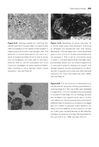
Figure 8.23 Histology section of a bird liver 40× Figure 8.24 Illustration of gross necropsy on
stained with Perls’ Prussian Blue iron stain to illus- a freshly dead aviary bird (parakeet) illustrating
trate the presence of iron stored as hemosiderin in an enlarged and discoloured liver with several
hepatocytes and Kupffer (macrophage) cells. The abscesses. Cultures taken from these abscesses
presence of excess hemosiderin can occur as a grew a pure culture of Yersinia pseudotuberculo-
result of excessive intake of iron or due to exces- sis serotype 2. This is not an uncommon cause
sive iron breakdown as in the case of haemolytic of death in wild and captive birds especially when
anaemia (that is, can be associated with avian predisposing factors such as immune-suppression
malaria) or subsequent to sever trauma and debili- or concurrent systemic disease are present. Iron
tation resulting in tissue damage and/or muscle storage diseases can also predispose to the devel-
breakdown. See also Plate 40. opment of pseudotuberculosis and other bacterial
infections (for more information see Cork, 2000).
See also Plate 41.
Figure 8.25 The use of immune-histochemistry to
identify lesions caused by Yersinia pseudotubercu-
losis (serotype 3) in the liver of the case illustrated
in Figure 8.24. Immuno-histochemistry is discussed
in Chapter 6. Essentially, the cut histology sections
are incubated with hyperimmune serum (in this
case rabbits anti-Yersinia pseudotuberculosis type 2
antisera) which is bound to an enzyme or conjugate
and then rinsed. A substrate is then added to the
slides and if the antibody remains bound to the cells
(or, in this case, bacterial lesions) on the slide this
indicates the presence of antigen (for more informa-
tion see Cork et al., 1999). See also Plate 42.
Vet Lab.indb 385 26/03/2019 10:26