
Page 417 - The Veterinary Laboratory and Field Manual 3rd Edition
P. 417
386 Susan C. Cork
Colville, T.P., Basset, J.M. (2007) Clinical Anatomy and
Physiology for Veterinary Technicians, 2nd edn.
Mosby, St Louis, MO.
Cork, S.C. (2000) Iron storage diseases in birds. Avian
Pathology 29(1): 7–12.
Cork, S.C., Collins-Emerson, J.M., Alley, M.R.,
Fenwick, S. (1999) Visceral lesions caused by
Yersinia pseudotuberculosis serotype 2 in different
species of bird. Avian Pathology 28(4) : 393–399.
Dyce, K.M., Sack, W.O., Wensing, C.J. (2009) Textbook
of Veterinary Anatomy, 4th edn. W.B. Saunders,
Philadelphia, PA.
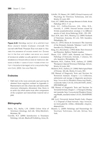
Figure 8.26 Histology section of a wild bird liver Getty, R. (1975) Sisson & Grossman’s The Anatomy
(New Zealand kokako [Callaeas cinereal]) 10× of the Domestic Animals, Volumes 1 and 2. W.B.
Saunders Co, Philadelphia, PA.
stained with Perls’ Prussian Blue iron stain to illus- King, A.S., McLelland, J. (1984) Birds: Their Structure
trate the presence of excess stored iron. Excess and Function. Bailliere Tindall, London.
iron in the liver and spleen can occur as a result Maxie, G.H. (2007) Jubb, Kennedy and Palmer’s
of excessive uptake (usually genetic) or excessive Pathology of Domestic Animals, 3 volumes, 5th
breakdown of blood cells as seen in haemolytic dis- edn. Saunders, Philadelphia, PA.
eases and also in cases of avian malaria where iron McGavin, M.D., Carlton, W.W., Zachary, J.F. (2000)
Thompson’s Special Veterinary Pathology, 3rd edn.
from infected and damaged cells is recycled at high Mosby, St Louis, MO.
rate (Cork, 2000). See also Plate 43. McGavin, M.D., Zachary, J.F. (2006) Pathological Basis
of Disease, 4th edn. Mosby, St Louis, MO.
OIE Manual of Diagnostic Tests and Vaccines for
Endnotes Terrestrial Animals. Chapter 1.1.2 Collection,
submission and storage of diagnostic specimens.
1 H&E stain is the most commonly used and may be http://www.oie.int/fileadmin/Home/eng/Health_
obtained from suppliers outlined in Appendix 4. standards/tahm/1.01.02_COLLECTION_DIAG_
Haematoxylin acts like a basic dye and stains acidic SPECIMENS.pdf.
structures (chromatin, ribosomes) blue. Eosin is OIE Manual of Diagnostic Tests and Vaccines for
an acidic dye which stains most other components Terrestrial Animals. Chapter 1.1.3 Transport of biolog-
of the cytoplasm and intercellular material red or ical materials. http://www.oie.int/fileadmin/Home/
pink. eng/Health_standards/tahm/1.01.03_TRANSPORT.
pdf.
OIE (n.d.) Terrestrial Animal Health Code. Chapter
4.12 Disposal of Dead Animals. http://www.oie.
Bibliography int/index.php?id=169&L=0&htmfile=chapitre_
disposal.htm.
Bacha, W.J., Bacha, L.M. (2000) Colour Atlas of Shapiro, L. (2009) Pathology and Parasitology for
Veterinary Histology, 2nd edn. Wiley-Blackwell, Veterinary Technicians, 2nd edn. Delmar, New
Hoboken, NJ. York.
Cheville, N.F. (2006) Introduction to Veterinary
Pathology, 3rd edn. Blackwell Publishing, Ames, IA.
Vet Lab.indb 386 26/03/2019 10:26