
Page 395 - Adams and Stashak's Lameness in Horses, 7th Edition
P. 395
Diagnostic Imaging 361
VetBooks.ir
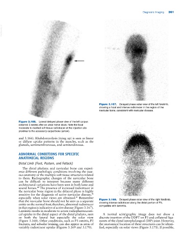
Figure 3.167. Delayed phase solar view of the left forelimb,
showing a focal and intense radiotracer in the region of the
navicular bone, consistent with navicular disease.
Figure 3.166. Lateral delayed phase view of the left carpus
obtained 3 weeks after an ulnar nerve block. Note the focal
moderate to marked soft tissue radiotracer at the injection site
proximal to the accessory carpal bone (arrow).
and 3.166). Rhabdomyolysis (tying up) is seen as linear
or diffuse uptake patterns in the muscles, such as the
gluteals, semimembranosus, and semitendinosus.
ABNORMAL CONDITIONS FOR SPECIFIC
ANATOMICAL REGIONS
Distal Limb (Foot, Pastern, and Fetlock)
The distal phalanx and navicular bone can experi
ence different pathologic conditions involving the osse
ous anatomy or the multiple soft tissue structures related
to them. Radiographic changes of the navicular bone
can be difficult to interpret because many different
architectural variations have been seen in both lame and
sound horses. The presence of increased radiotracer in
44
the navicular bone region in the delayed phase is highly
sensitive for the diagnosis of active navicular disease,
92
especially when solar views are obtained. Remember
45
that the navicular bone should not be seen as a separate Figure 3.168. Delayed phase solar view of the right hindlimb,
entity on the normal foot; therefore, abnormal radiotracer showing intense radiotracer along the distal portion of P3,
in that region is indicative of active disease (Figure 3.167). compatible with laminitis.
Laminitis results in moderate to severe radiopharmaceuti
cal uptake in the distal aspect of the distal phalanx, seen A normal scintigraphic image does not show a
on both the lateral but especially the solar view discrete insertion of the DDFT on P3 and collateral liga
(Figure 3.168). Other conditions, such as P3 osteitis, P3 ments of the distal interphalangeal (DIP) joint. However,
fractures, and subsolar bruising, can also appear as areas of the anatomical location of these structures can be identi
variably radiotracer uptake (Figures 3.169 and 3.170). fied, especially on solar views (Figure 3.171). If possible,