
Page 399 - Adams and Stashak's Lameness in Horses, 7th Edition
P. 399
Diagnostic Imaging 365
VetBooks.ir
A B
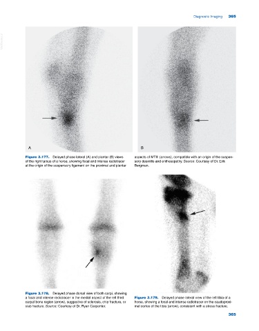
Figure 3.177. Delayed phase lateral (A) and plantar (B) views aspects of MTIII (arrows), compatible with an origin of the suspen
of the right tarsus of a horse, showing focal and intense radiotracer sory desmitis and enthesopathy. Source: Courtesy of Dr. Erik
at the origin of the suspensory ligament on the proximal and plantar Bergman.
Figure 3.178. Delayed phase dorsal view of both carpi, showing
a focal and intense radiotracer in the medial aspect of the left third Figure 3.179. Delayed phase lateral view of the left tibia of a
carpal bone region (arrow), suggestive of sclerosis, chip fracture, or horse, showing a focal and intense radiotracer on the caudoproxi
slab fracture. Source: Courtesy of Dr. Ryan Carpenter. mal cortex of the tibia (arrow), consistent with a stress fracture.
365