
Page 397 - Adams and Stashak's Lameness in Horses, 7th Edition
P. 397
Diagnostic Imaging 363
VetBooks.ir
Figure 3.171. (A) Delayed phase solar view of a normal horse.
Note the uniform uptake along the entire image. NB = navicular
bone area and DDFT = deep digital flexor tendon area. (B) SHINE‐
processed image.
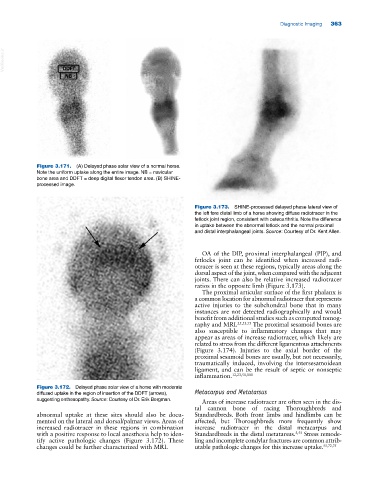
Figure 3.173. SHINE‐processed delayed phase lateral view of
the left fore distal limb of a horse showing diffuse radiotracer in the
fetlock joint region, consistent with osteoarthritis. Note the difference
in uptake between the abnormal fetlock and the normal proximal
and distal interphalangeal joints. Source: Courtesy of Dr. Kent Allen.
OA of the DIP, proximal interphalangeal (PIP), and
fetlocks joint can be identified when increased radi
otracer is seen at these regions, typically areas along the
dorsal aspect of the joint, when compared with the adjacent
joints. There can also be relative increased radiotracer
ratios in the opposite limb (Figure 3.173).
The proximal articular surface of the first phalanx is
a common location for abnormal radiotracer that represents
active injuries to the subchondral bone that in many
instances are not detected radiographically and would
benefit from additional studies such as computed tomog
raphy and MRI. 25,53,73 The proximal sesamoid bones are
also susceptible to inflammatory changes that may
appear as areas of increase radiotracer, which likely are
related to stress from the different ligamentous attachments
(Figure 3.174). Injuries to the axial border of the
proximal sesamoid bones are usually, but not necessarily,
traumatically induced, involving the intersesamoidean
ligament, and can be the result of septic or nonseptic
inflammation. 12,23,51,101
Figure 3.172. Delayed phase solar view of a horse with moderate
diffused uptake in the region of insertion of the DDFT (arrows), Metacarpus and Metatarsus
suggesting enthesopathy. Source: Courtesy of Dr. Erik Bergman.
Areas of increase radiotracer are often seen in the dis
tal cannon bone of racing Thoroughbreds and
abnormal uptake at these sites should also be docu Standardbreds. Both front limbs and hindlimbs can be
mented on the lateral and dorsal/palmar views. Areas of affected, but Thoroughbreds more frequently show
increased radiotracer in these regions in combination increase radiotracer in the distal metacarpus and
with a positive response to local anesthesia help to iden Standardbreds in the distal metatarsus. 5,55 Stress remode
tify active pathologic changes (Figure 3.172). These ling and incomplete condylar fractures are common attrib
changes could be further characterized with MRI. utable pathologic changes for this increase uptake. 55,72,75