
Page 401 - Adams and Stashak's Lameness in Horses, 7th Edition
P. 401
Diagnostic Imaging 367
VetBooks.ir
A B
C
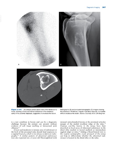
Figure 3.181. (A) Delayed phase lateral view of the left tibia of a Radiographic (B) and computed tomographic (C) images showing
horse, showing a focal and intense radiotracer in the medullary the presence of ill‐defined, irregular medullary sclerosis, compatible
cavity of the proximal diaphysis, suggestive of enostosis‐like lesion. with an enostosis‐like lesion. Source: Courtesy of Dr. Erik Bergman.
is a rare condition in horses and can be a diagnostic unusual osteochondral lesions at the proximal articular
76
challenge because the patient can present without margin of the medial trochlear ridge of the talus.
evidence of soft tissue swelling or tarsocrural joint Osseous cyst‐like lesions in the tarsus have been
effusion. 16 described as focal areas of abnormal radiotracer in the
A focal and moderate to intense area of radiotracer at distal tibia (medial or lateral malleoli or intermediate
36
the level of the proximal talus should lend suspicion to sagittal ridge) or talus. Adding a flexed lateral view to
a sagittal fracture, which is not always evident on radio the routine scintigraphic exam protocol of the tarsus
graphs. A similar pattern of abnormal radiotracer can help to differentiate whether the abnormal radi
16
was also reported on a 2‐year‐old Thoroughbred with otracer is located in the distal tibia or the talus.