
Page 403 - Adams and Stashak's Lameness in Horses, 7th Edition
P. 403
VetBooks.ir
A B
C
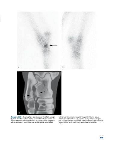
Figure 3.184. Delayed phase lateral views of the left (A) and right right tarsus. Computed tomographic image (C) of the left tarsus
(B) tarsi. Note the focal and intense abnormal radiotracer seen in the confirms the osteoarthritis with evidence of irregular bone margins
region of the talocalcaneal joint on the left tarsus (arrow), compatible with adjacent sclerosis and soft tissue mineralization of the intertarsal
with osteoarthritis. Compare with the uniform uptake of the normal region (arrows). Source: Courtesy of Dr. Sarah M. Puchalski.
369