
Page 404 - Adams and Stashak's Lameness in Horses, 7th Edition
P. 404
VetBooks.ir
Figure 3.186. Delayed phase tail on detector (TOD) view of the
pelvis of a horse, showing focal and intense increased radiotracer
on the right ischial tuberosity, compatible with a fracture. Source:
Courtesy of Dr. Erik Bergman.
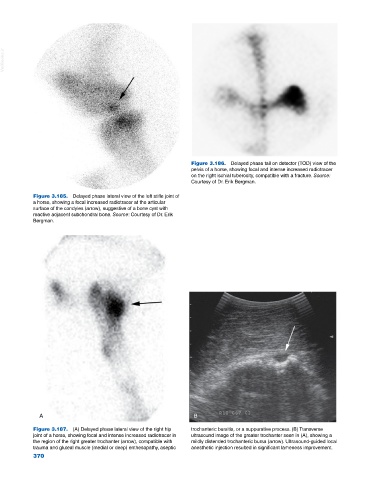
Figure 3.185. Delayed phase lateral view of the left stifle joint of
a horse, showing a focal increased radiotracer at the articular
surface of the condyles (arrow), suggestive of a bone cyst with
reactive adjacent subchondral bone. Source: Courtesy of Dr. Erik
Bergman.
A B
Figure 3.187. (A) Delayed phase lateral view of the right hip trochanteric bursitis, or a suppurative process. (B) Transverse
joint of a horse, showing focal and intense increased radiotracer in ultrasound image of the greater trochanter seen in (A), showing a
the region of the right greater trochanter (arrow), compatible with mildly distended trochanteric bursa (arrow). Ultrasound‐guided local
trauma and gluteal muscle (medial or deep) enthesopathy, aseptic anesthetic injection resulted in significant lameness improvement.
370