
Page 407 - Adams and Stashak's Lameness in Horses, 7th Edition
P. 407
Diagnostic Imaging 373
In summary, the practitioner should be aware of the
potential limitations when interpreting the results of a
VetBooks.ir the signalment, history, clinical signs, and physical/lame
nuclear scintigraphic examination; correlate them with
ness exam findings; and consider further diagnostics
when appropriate.
References
1. Allhands RV, Twardock AR, Boero MJ. Uptake of 99m Tc‐MDP in
muscle associated with peripheral nerve block. Vet Radiol
Ultrasound 1987;28:181–184.
2. Anderson JD, Galuppo LD, Barr BC, et al. Clinical and scinti
graphic findings in horses with a bone fragility disorder: 16 cases
(1980–2006). J Am Vet Med Assoc 2008;232:1694–1699.
3. Archer DC, Boswell JC, Voute LC, et al. Skeletal scintigraphy in
the horse: current indications and validity as a diagnostic test. Vet
J 2007;173:31–44.
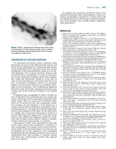
Figure 3.193. Delayed phase left lateral view of the caudal 4. Arndt J, Pauwels E, Camps J, et al. Clinical differences between
cervical region of a horse, showing marked, focal, and intense bone‐seeking agents. Eur J Nucl Med 1985;11:330.
abnormal radiotracer at the articular facets of C6–C7 (arrow), 5. Arthur RM, Constantinide D. Results of 428 nuclear scintigraphic
compatible with osteoarthritis. examinations of the musculoskeletal system at a Thoroughbred
racetrack. Proc Am Assoc Equine Pract 1995;41:84–87.
6. Bailey RE, Dyson SJ, Parkin TD. Focal increased radiopharmaceu
tical uptake in the dorsoproximal diaphyseal region of the equine
LIMITATIONS OF NUCLEAR MEDICINE proximal phalanx. Vet Radiol Ultrasound 2007;48:460–466.
7. Bassage LH, Ross MW. Enostosis‐like lesions in the long bones of
As mentioned previously, nuclear medicine traces 10 horses: scintigraphic and radiographic features. Equine Vet J
physiologic processes in the different body systems and 1998;30:35–42.
therefore has a high sensitivity in detecting early changes 8. Berry CR, Daniel GB. Pulmonary and mucociliary scintigraphy. In
Textbook of Veterinary Nuclear Medicine, 2nd ed. Daniel GB,
in the metabolism. This is the main reason why nuclear Berry CR, eds. North Carolina State University Press, Raleigh,
scintigraphy of the musculoskeletal system is an invalu NC, 2006;303–327.
able diagnostic tool in equine lameness. However, the 9. Butson RJ, Webbon PM, Fairbairn SM. 99m Tc‐HMPAO labeled
specificity of bone scintigraphy is very low in the major leucocytes and their biodistribution in the horse: a preliminary
investigation. Equine Vet J 1995;27:313–315.
ity of cases. During the evaluation of the bone scintigra 10. Chambers MD, Martinelli MJ, Baker GJ, et al. Nuclear medicine
phy results, the practitioner should be aware that an for diagnosis of lameness in horses. J Am Vet Med Assoc
area of increased radiotracer only means that there is an 1995;206:792–796.
area of increased osteoblastic activity in a particular 11. Dabareiner RM, Cole RC. Fractures of the tuber coxae of the
region. If the increased radiotracer is considered patho ilium in horses: 29 cases (1996–2007). J Am Vet Med Assoc
2009;234:1303–1307.
logic, in most situations, a list of differential diagnoses 12. Dabareiner RM, Watkins JP, Carter GK, et al. Osteitis of the axial
should be made and further imaging performed to gain border of the proximal sesamoid bones in horses: eight cases
a better idea of the anatomical changes of the affected (1993–1999). J Am Vet Med Assoc 2001;219:82–86.
region. 13. Davenport‐Goodall CLM, Ross MW. Scintigraphic abnormalities
of the pelvic region in horses examined because of lameness or
In general, bone scintigraphy is used to localize an poor performance: 128 cases (1993–2000). J Am Vet Med Assoc
area of abnormal bone metabolism that may explain the 2004;224:88–95.
source of lameness and not necessarily the specific path 14. David GW, Elizabeth R. Nonsurgical management of ulnar frac
ologic change. There are some cases in which the diag tures in the horse: a retrospective study of 43 cases. Vet Surg
1985;14:283–286.
nosis can be made from the bone scintigraphy results. 15. Davidson EJ, Martin BB, Jr. Stress fracture of the scapula in two
For example, one can diagnose a stress fracture in a horses. Vet Radiol Ultrasound 2004;45:407–410.
sound racehorse that becomes lame immediately after a 16. Davidson EJ, Ross MW, Parente EJ. Incomplete sagittal fracture
training session or a race and has a focal increased radi of the talus in 11 racehorses: outcome. Equine Vet J 2005;
37:457–461.
otracer in the diaphysis of the tibia. On the other hand, 17. Denoix JM. Discovertebral pathology in horses. Equine Vet Educ
if a similar area of increased radiotracer is found in a 2007;19:72–73.
retired older horse that spends most of the time in the 18. Duggan VE, Holbrook TC, Dechant JE, et al. Diagnosis of aortoil
pasture, other differential diagnoses should be consid iac thrombosis in a Quarter horse foal using Doppler ultrasound
and nuclear scintigraphy. J Vet Intern Med 2004;18:753–756.
ered, such as blunt trauma with or without a fracture, 19. Dyson S. Sixteen fractures of the shoulder region in the horse.
enostosis‐like lesion, osteomyelitis, or neoplasia. Equine Vet J 1985;17:104–110.
False‐negative results occur secondary to many dif 20. Dyson S. Shoulder lameness in horses: an analysis of 58 suspected
ferent reasons and can be considered as a limitation of cases. Equine Vet J 1986;18:29–36.
bone scintigraphy. For example, a bone lesion with min 21. Dyson SJ. Subjective and quantitative scintigraphic assessment of
the equine foot and its relationship with foot pain. Equine Vet J
imum uptake in the proximal region of a limb may not 2002;34:164–170.
be apparent due to the significant γ‐ray attenuation 22. Dyson S, Murray R. Pain associated with the sacroiliac joint
produced by the surrounding musculature. As in cases of region: a clinical study of 74 horses. Equine Vet J 2003;
35:240–245.
subchondral bone cysts, cartilage or meniscal injuries 23. Dyson SJ, Murray R. Osseous trauma in the fetlock region of
will likely give a false‐negative result if the adjacent mature sport horses. Proc. Am Assoc Equine Pract 2006;
bone is not affected. 52:443–456.