
Page 406 - Adams and Stashak's Lameness in Horses, 7th Edition
P. 406
372 Chapter 3
VetBooks.ir
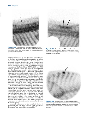
Figure 3.190. Delayed phase LDO view of the mid‐thoracic
vertebrae of a normal horse. Note the relative increased uptake of Figure 3.191. Delayed phase LDO view of the mid‐thoracic
the vertebral bodies when compared with the corresponding dorsal vertebrae of a horse, showing focal and intense abnormal radi
spinous processes (arrows). otracer at least two adjacent dorsal spinous processes (arrows),
suggestive of impingement.
the lumbar spine, can be very difficult to obtain because
of the large amount of musculature causing considera
ble scatter radiation and because if the technique is
increased, the most dorsal aspect of the dorsal spinous
processes can be overexposed. This makes the scinti
graphic evaluation of the spine an invaluable tool for
evaluation of the back (Figure 3.190). LDO and RDO
views of the spine are the best, although straight lateral
and dorsal views are made if a lesion is found. Areas of
mild abnormal radiotracer at the dorsal aspect of the
spinous processes can be seen in horses with no clinical
signs of back problems; therefore, these findings should
be interpreted with caution. 33,34 Focal and intense areas
of radiotracer can be seen with fractures, severe impinge
ment with or without ligamentous damage, osteomyeli
tis, or neoplasia (Figures 3.191 and 3.192). Diagnostic
differentials should be made, taking into consideration
history, physical/clinical exam, and blood analysis.
Increased radiotracer in the cervical articular facets is
most commonly seen in cases of OA. The increased radi
otracer can be identified when the bone activity in the
region of the articular facets is greater than in the cor
responding vertebral body or adjacent joints. Affected
cervical vertebral facets may be unilateral or bilateral,
and more than one joint may be affected (Figure 3.193).
Therefore, left and right views should be obtained, and
the practitioner should be aware of the possibility of
multiple adjacent sites affected and not rely only on the
comparison of adjacent joints for increased radiotracer Figure 3.192. Delayed phase LDO view of the withers of a
in the evaluation. horse, showing a focal and intense abnormal radiotracer involving
Increased radiotracer in the vertebral bodies is only the most proximal aspect of one of the spinous processes
uncommon and has been reported with spondylosis (arrow), suggestive of a fracture. Source: Courtesy of Dr. Erik
deformans and cases of discospondylitis. 40,85 Bergman.
17