
Page 419 - Adams and Stashak's Lameness in Horses, 7th Edition
P. 419
VetBooks.ir
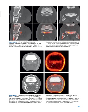
Figure 3.204. 18F‐NaF PET CT of mild navicular There are no significant abnormalities of the navicular bone on the
remodeling.Multiplanar reformatted CT (top) and fused 18F‐NaF CT images, but the PET demonstrates marked focal uptake of the
PET/CT (bottom) images of the left front foot of a 10‐year‐old flexor cortex of the navicular bone, worse medially. (Arrow) This
Warmblood with lameness localized to the foot. Medial is to the left. indicates early remodeling of the navicular bone.
Figure 3.205. Deep digital flexor tendon lesion imaged with pre‐contrast CT (arrowheads). There is marked focal 18F‐FDG
18F‐FDG PET/CT.Transverse CT (top left), 18F‐FDG PET (top uptake of the lateral lobe of the tendon (upper right arrow). Arterial
right), arterial contrast CT (bottom left), and fused PET/CT (bottom contrast enhancement is also present on CT. Note that on arterial
right) images of the right front foot of a 22‐year‐old Quarter Horse contrast CT, both the lesions and adjacent vessels demonstrate
mare through the middle phalanx. Lateral is to the left. The lateral enhancement(lower left arrow), whereas on the PET images only
lobe of the DDFT appears enlarged with irregular margin on the the lesion shows increased uptake (lower right arrow).
385