
Page 416 - Adams and Stashak's Lameness in Horses, 7th Edition
P. 416
382 Chapter 3
VetBooks.ir
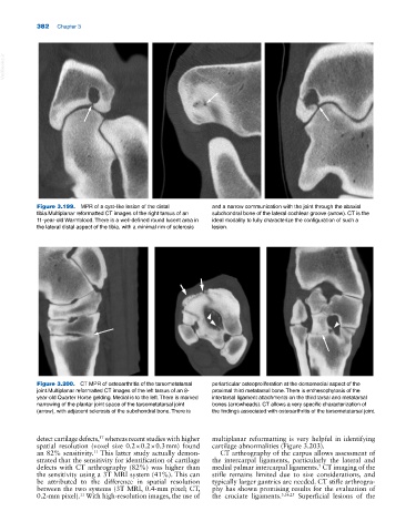
Figure 3.199. MPR of a cyst‐like lesion of the distal and a narrow communication with the joint through the abaxial
tibia.Multiplanar reformatted CT images of the right tarsus of an subchondral bone of the lateral cochlear groove (arrow). CT is the
11‐year‐old Warmblood. There is a well‐defined round lucent area in ideal modality to fully characterize the configuration of such a
the lateral distal aspect of the tibia, with a minimal rim of sclerosis lesion.
Figure 3.200. CT MPR of osteoarthritis of the tarsometatarsal periarticular osteoproliferation at the dorsomedial aspect of the
joint.Multiplanar reformatted CT images of the left tarsus of an 8‐ proximal third metatarsal bone. There is enthesophytosis of the
year‐old Quarter Horse gelding. Medial is to the left. There is marked intertarsal ligament attachments on the third tarsal and metatarsal
narrowing of the plantar joint space of the tarsometatarsal joint bones (arrowheads). CT allows a very specific characterization of
(arrow), with adjacent sclerosis of the subchondral bone. There is the findings associated with osteoarthritis of the tarsometatarsal joint.
detect cartilage defects, whereas recent studies with higher multiplanar reformatting is very helpful in identifying
17
spatial resolution (voxel size 0.2 × 0.2 × 0.3 mm) found cartilage abnormalities (Figure 3.203).
an 82% sensitivity. This latter study actually demon CT arthrography of the carpus allows assessment of
11
strated that the sensitivity for identification of cartilage the intercarpal ligaments, particularly the lateral and
defects with CT arthrography (82%) was higher than medial palmar intercarpal ligaments. CT imaging of the
7
the sensitivity using a 3T MRI system (41%). This can stifle remains limited due to size considerations, and
be attributed to the difference in spatial resolution typically larger gantries are needed. CT stifle arthrogra
between the two systems (3T MRI, 0.4‐mm pixel; CT, phy has shown promising results for the evaluation of
0.2‐mm pixel). With high‐resolution images, the use of the cruciate ligaments. 5,16,25 Superficial lesions of the
11