
Page 434 - Adams and Stashak's Lameness in Horses, 7th Edition
P. 434
400 Chapter 3
VetBooks.ir
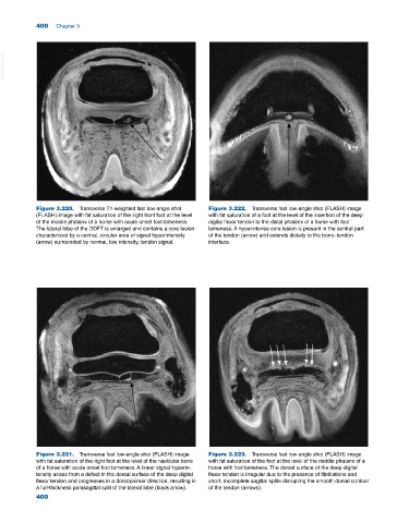
Figure 3.220. Transverse T1‐weighted fast low‐angle shot Figure 3.222. Transverse fast low‐angle shot (FLASH) image
(FLASH) image with fat saturation of the right front foot at the level with fat saturation of a foot at the level of the insertion of the deep
of the middle phalanx of a horse with acute onset foot lameness. digital flexor tendon to the distal phalanx of a horse with foot
The lateral lobe of the DDFT is enlarged and contains a core lesion lameness. A hyperintense core lesion is present in the central part
characterized by a central, circular area of signal hyperintensity of the tendon (arrow) and extends distally to the bone–tendon
(arrow) surrounded by normal, low intensity, tendon signal. interface.
Figure 3.221. Transverse fast low‐angle shot (FLASH) image Figure 3.223. Transverse fast low‐angle shot (FLASH) image
with fat saturation of the right foot at the level of the navicular bone with fat saturation of the foot at the level of the middle phalanx of a
of a horse with acute onset foot lameness. A linear signal hyperin horse with foot lameness. The dorsal surface of the deep digital
tensity arises from a defect in the dorsal surface of the deep digital flexor tendon is irregular due to the presence of fibrillations and
flexor tendon and progresses in a dorsopalmar direction, resulting in short, incomplete sagittal splits disrupting the smooth dorsal contour
a full‐thickness parasagittal split of the lateral lobe (black arrow). of the tendon (arrows).
400