
Page 435 - Adams and Stashak's Lameness in Horses, 7th Edition
P. 435
Diagnostic Imaging 401
Lesions of the Navicular Bone concurrent abnormalities of the distal border or flexor
surface on MR images. In such horses, it is difficult to
MR images of the navicular bone in horses with
VetBooks.ir navicular bone disease may show one or more of three differentiate between abnormal osseous fluid of an acute
post‐traumatic nature and osseous fluid accumulation
main abnormalities: remodeling changes in the medulla,
associated with a chronic degenerative process on a sin
degenerative changes of the flexor border, and osteo
chondral fragmentation of the distal border. The most gle examination. As acute, post‐traumatic osseous fluid
49
(e.g. a bone contusion) tends to resolve with rest, in con
common type of abnormality seen in the navicular bones
of horses with recent onset navicular syndrome was trast to the persistence of degenerative osseous fluid, this
distinction requires one or more follow‐up MRI exami
STIR signal hyperintensity in the medullary cavity of the 190
navicular bone with or without additional areas of T2 nations to be performed.
Medullary STIR signal hyperintensity may be focal
and PD signal hypointensity (Figure 3.224), but this
152
STIR signal increase is more reliably assessed on FSE near the distal or palmar border of the navicular bone, or
extend from the distal border in a vertical band along the
than on GRE images. It has been speculated that osse
138
ous fluid in the medulla may be an acute inflammatory palmar cortex to the proximal border of the bone, or
spread diffusely throughout the medullary cavity. Based
or post‐traumatic finding in horses with recent onset
navicular syndrome. However, in pathological stud on the extent and intensity of STIR signal increase, med
152
ullary bone edema can be graded from mild to severe.
ies, MRI evidence of “bone marrow lesion” was associ
ated not only with acute inflammation but also with Lower grades of medullary STIR signal hyperintensity
have been encountered in non‐lame control limbs, but
evidence of hemorrhage, interstitial edema in medullary
fat, prominent capillary infiltration in marrow fat, thin severe medullary edema was strongly associated with the
presence of lameness.
Areas of signal hypointensity
121,152
ning of trabeculae and widening of intertrabecular
spaces, medullary fibrosis, chronic osteonecrosis, and, in may also be seen in the medullary cavity of the navicular
bone and may be focal or diffuse. Loss of T1 signal may
some horses, adipose tissue necrosis. 17,32,58 In one patho
logical study, all but one horse with medullary STIR sig either represent replacement of medullary fat by fluid or
bone densification, while loss of T2 and PD signal indi
nal hyperintensity had concurrent chronic degenerative
changes of the flexor surface of the navicular bone. cates the presence of bone densification and sclerosis or
158
even replacement of medullary trabecular bone by com
On the other hand, pathological studies have not
included horses with recent onset lameness, and it there pact lamellar bone, usually as a consequence of marked
trabecular thickening in response to degenerative changes
fore remains possible that medullary STIR hyperinten 4,158
sity can represent acute inflammatory fluid in the of the flexor surface of the navicular bone.
In horses with chronic navicular syndrome, the most
152
spongiosa of the navicular bone. This possibility is
supported by the frequent observation of osseous fluid common MRI abnormality was the presence of abnor
mal signal hyperintensity at the level of the flexor bor
signal in the spongiosa of the navicular bone without
der of the navicular bone (Figure 3.225). This can be
158
a subtle, focal increase, caused by synovial fluid pooling
at a site of palmar fibrocartilage loss and thinning, best
seen on sagittal PD or T2‐weighted images. MRI bur
sography with saline has been shown to improve the
conspicuity of fibrocartilage lesions. 159
A normal shallow, smooth depression is present in
the middle third of the palmar sagittal ridge of up to
197
50% of normal navicular bones. This is also charac
terized by pooling of synovial bursal fluid but should
not be confused with a degenerative lesion of the palmar
fibrocartilage. Even so, this normal synovial depression
may be the site of early bone degeneration in some
horses with navicular bone disease. The presence of
osseous fluid signal on STIR images within the flexor
cortex and spongiosa adjacent to the synovial depres
sion in the sagittal ridge is a likely sign of early bone
degeneration. Signal increase at the flexor surface can
also be more extensive and extend deeper within the
cortical bone of the flexor cortex when cortical bone
erosion is present (Figure 3.225). Focal bone loss from
the flexor surface is best seen on fat‐suppressed images
in high‐field studies and transverse high‐resolution T1
images on low‐field standing MR images. These flexor
cortex lesions may not be easily detected radiographi
cally. In affected horses, MR images usually show
165
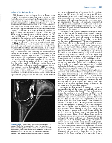
Figure 3.224. Sagittal short tau inversion recovery (STIR)
image of the foot of a horse with lameness that is abolished by concurrent irregularity of the normally smooth endosteal
anesthesia of the palmar digital nerves. There is marked STIR surface of the flexor cortex, especially opposite the site
hyperintensity of cancellous bone in the medullary cavity of the of focal signal increase of the palmar surface.
navicular bone (arrow) indicating the presence of abnormal Fibrocartilage and cortical bone loss at the palmar
medullary fluid, medullary fibrosis, or medullary fat necrosis. aspect of the flexor surface of the navicular bone is