
Page 440 - Adams and Stashak's Lameness in Horses, 7th Edition
P. 440
406 Chapter 3
articular cartilage layer. The resulting narrowing of the margin and disruption of the adjacent laminar architecture.
joint space width may be an early sign of osteoarthri Chronic bone mineral density increase of a palmar pro
VetBooks.ir bearing weight symmetrically in a low‐field magnet or bone bruise, or may be associated with collateral liga
cess of the distal phalanx may be the result of a chronic
tis, but this sign is only reliable if the horse is either
149
ment injury. Bone bruising of the dorsodistal aspect of
the foot is supported with the limb held straight in a
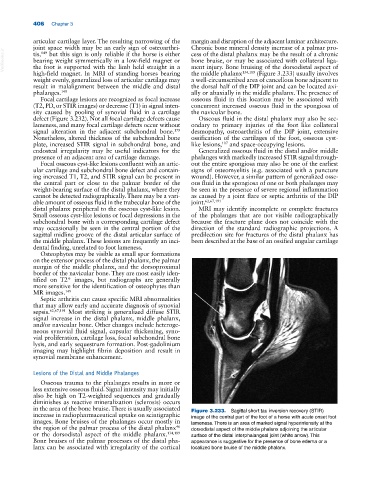
high‐field magnet. In MRI of standing horses bearing the middle phalanx 134,189 (Figure 3.233) usually involves
weight evenly, generalized loss of articular cartilage may a well‐circumscribed area of cancellous bone adjacent to
result in malalignment between the middle and distal the dorsal half of the DIP joint and can be located axi
phalanges. 149 ally or abaxially in the middle phalanx. The presence of
Focal cartilage lesions are recognized as focal increase osseous fluid in this location may be associated with
(T2, PD, or STIR images) or decrease (T1) in signal inten concurrent increased osseous fluid in the spongiosa of
sity caused by pooling of synovial fluid in a cartilage the navicular bone.
defect (Figure 3.232). Not all focal cartilage defects cause Osseous fluid in the distal phalanx may also be sec
lameness, and many focal cartilage defects occur without ondary to primary injuries of the foot like collateral
signal alteration in the adjacent subchondral bone. desmopathy, osteoarthritis of the DIP joint, extensive
179
Nonetheless, altered thickness of the subchondral bone ossification of the cartilages of the foot, osseous cyst‐
plate, increased STIR signal in subchondral bone, and like lesions, and space‐occupying lesions.
107
endosteal irregularity may be useful indicators for the Generalized osseous fluid in the distal and/or middle
presence of an adjacent area of cartilage damage. phalanges with markedly increased STIR signal through
Focal osseous cyst‐like lesions confluent with an artic out the entire spongiosa may also be one of the earliest
ular cartilage and subchondral bone defect and contain signs of osteomyelitis (e.g. associated with a puncture
ing increased T1, T2, and STIR signal can be present in wound). However, a similar pattern of generalized osse
the central part or close to the palmar border of the ous fluid in the spongiosa of one or both phalanges may
weight‐bearing surface of the distal phalanx, where they be seen in the presence of severe regional inflammation
cannot be detected radiographically. There may be a vari as caused by a joint flare or septic arthritis of the DIP
able amount of osseous fluid in the trabecular bone of the joint. 62,67,191
distal phalanx peripheral to the osseous cyst‐like lesion. MRI may identify incomplete or complete fractures
Small osseous cyst‐like lesions or focal depressions in the of the phalanges that are not visible radiographically
subchondral bone with a corresponding cartilage defect because the fracture plane does not coincide with the
may occasionally be seen in the central portion of the direction of the standard radiographic projections. A
sagittal midline groove of the distal articular surface of predilection site for fractures of the distal phalanx has
the middle phalanx. These lesions are frequently an inci been described at the base of an ossified ungular cartilage
dental finding, unrelated to foot lameness.
Osteophytes may be visible as small spur formations
on the extensor process of the distal phalanx, the palmar
margin of the middle phalanx, and the dorsoproximal
border of the navicular bone. They are most easily iden
tified on T2* images, but radiographs are generally
more sensitive for the identification of osteophytes than
MR images. 149
Septic arthritis can cause specific MRI abnormalities
that may allow early and accurate diagnosis of synovial
sepsis. 62,67,191 Most striking is generalized diffuse STIR
signal increase in the distal phalanx, middle phalanx,
and/or navicular bone. Other changes include heteroge
neous synovial fluid signal, capsular thickening, syno
vial proliferation, cartilage loss, focal subchondral bone
lysis, and early sequestrum formation. Post‐gadolinium
imaging may highlight fibrin deposition and result in
synovial membrane enhancement.
Lesions of the Distal and Middle Phalanges
Osseous trauma to the phalanges results in more or
less extensive osseous fluid. Signal intensity may initially
also be high on T2‐weighted sequences and gradually
diminishes as reactive mineralization (sclerosis) occurs
in the area of the bone bruise. There is usually associated Figure 3.233. Sagittal short tau inversion recovery (STIR)
increase in radiopharmaceutical uptake on scintigraphic image of the central part of the foot of a horse with acute onset foot
images. Bone bruises of the phalanges occur mostly in lameness. There is an area of marked signal hyperintensity at the
the region of the palmar process of the distal phalanx dorsodistal aspect of the middle phalanx adjoining the articular
46
or the dorsodistal aspect of the middle phalanx. 134,189 surface of the distal interphalangeal joint (white arrow). This
Bone bruises of the palmar processes of the distal pha appearance is suggestive for the presence of bone edema or a
lanx can be associated with irregularity of the cortical localized bone bruise of the middle phalanx.