
Page 443 - Adams and Stashak's Lameness in Horses, 7th Edition
P. 443
Diagnostic Imaging 409
MAGNETIC RESONANCE IMAGING abaxial sesamoid nerve block in the majority of
but also, though less frequently, by a palmar
horses
OF THE FETLOCK REGION
35,51
VetBooks.ir Introduction digital nerve block. Unfortunately, this regularly causes
37
confusion as to which region of the distal limb should be
evaluated first with MRI in horses that are presented
Conventional imaging techniques have limitations in
the fetlock region. Radiography and scintigraphy are with a history of lameness resolution following either of
not capable of detecting early cartilage loss and sub these nerve blocks. Experience with such patients teaches
chondral bone injury without marked structural bone that horses presented with a history of a negative
damage or demineralization. 54,166,202 Ultrasonographic response to a palmar digital nerve block but a positive
evaluation lacks sensitivity for injuries of the suspensory response to an abaxial sesamoid nerve block are much
branches and the straight and oblique distal sesamoid more likely to have a primary fetlock lesion than a foot
35
ean ligaments. 152,172 Hence MRI is being used routinely lesion responsible for the lameness.
to diagnose the causes of lameness in the fetlock region The normal MRI anatomy of the fetlock joint has
of horses. The primary indication for MRI is pain local been described, using both high‐ and low‐field images
173
ized to the fetlock region with diagnostic analgesia, (MRI abnormalities in the fetlock region). Several
without sufficient radiological or ultrasonographic studies have documented the incidence of MRI diagno
70,90,144
abnormalities to explain the degree of lameness. The ses in the fetlock region (Table 3.4). Types and
increasingly frequent use of MRI to elucidate distal limb incidence of lesions in the fetlock differ depending on
lameness has revealed that lameness caused by pain in horse breed and athletic activity. In a study of 40 horses
the fetlock region may be abolished not only by an from a variety of breeds and disciplines, half the horses
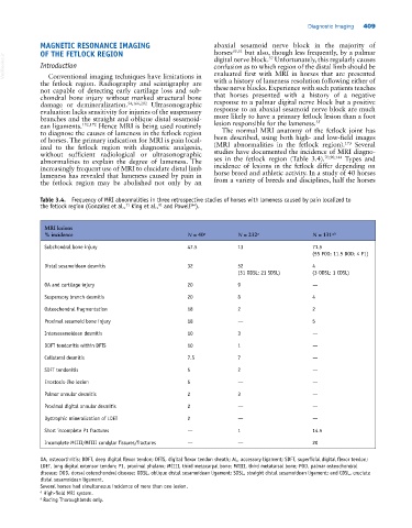
Table 3.4. Frequency of MRI abnormalities in three retrospective studies of horses with lameness caused by pain localized to
the fetlock region (Gonzalez et al., King et al., and Powell ).
144
91
71
MRI lesions
% incidence N = 40 a N = 232 a N = 131 a,b
Subchondral bone injury 47.5 13 71.5
(55 POD; 11.5 DOD; 4 P1)
Distal sesamoidean desmitis 32 52 4
(31 ODSL; 21 SDSL) (3 ODSL; 1 CDSL)
OA and cartilage injury 20 9 —
Suspensory branch desmitis 20 8 4
Osteochondral fragmentation 18 2 2
Proximal sesamoid bone injury 18 — 5
Intersesamoidean desmitis 10 3 —
DDFT tendonitis within DFTS 10 1 —
Collateral desmitis 7.5 7 —
SDFT tendonitis 5 2 —
Enostosis‐like lesion 5 — —
Palmar annular desmitis 2 3 —
Proximal digital annular desmitis 2 — —
Dystrophic mineralization of LDET 2 — —
Short incomplete P1 fractures — 1 14.5
Incomplete MCIII/MTIII condylar fissures/fractures — — 20
OA, osteoarthritis; DDFT, deep digital flexor tendon; DFTS, digital flexor tendon sheath; AL, accessory ligament; SDFT, superficial digital flexor tendon;
LDET, long digital extensor tendon; P1, proximal phalanx; MCIII, third metacarpal bone; MTIII, third metatarsal bone; POD, palmar osteochondral
disease; DOD, dorsal osteochondral disease; ODSL, oblique distal sesamoidean ligament; SDSL, straight distal sesamoidean ligament; and CDSL, cruciate
distal sesamoidean ligament.
Several horses had simultaneous incidence of more than one lesion.
a High‐field MRI system.
b Racing Thoroughbreds only.