
Page 445 - Adams and Stashak's Lameness in Horses, 7th Edition
P. 445
Diagnostic Imaging 411
extensive osseous fluid are more likely to be clinically
significant than lower‐grade lesions in this location.
VetBooks.ir intensity on T2 and STIR images and decreased signal
Normal, parallel, linear areas of increased signal
intensity on T1 images may be present in the palmar
region of the distal metaphysis and epiphysis of the
MCIII/MTIII. This “tiger‐stripe” appearance is due to
hypervascularity or congestion of the nutrient vessels
that appear as well‐defined hyperintense lines emanat
ing from the palmar aspect of the metaphyseal region
coursing in a dorsodistal direction into the condyles of
the MCIII/MTIII. An increase in number, size, and con
spicuity of these vessels may occur in the presence of
chronic remodeling/sclerosis of the distal condyles or
capsulitis with distension of the joint.
The ability of MRI to predict which horses are at risk
of developing complete condylar or proximal sesamoid
bone fractures has been studied extensively. 140,141,147,182,183
Despite conflicting information on a clear association
between sclerosis and the ability to predict condylar
fracture formation, there was an overall higher grade of
osseous fluid (bone marrow lesion) and bone densifica
tion (sclerosis) in the condyle of bones with a condylar
fracture, than in the condyles of the limbs of control
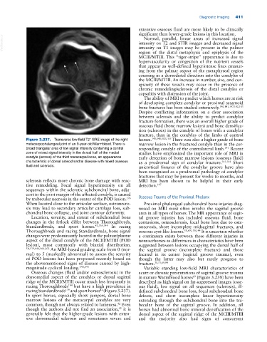
Figure 3.237. Transverse low‐field T2* GRE image of the right horses. 140,141,182,183 There was also a higher grade of bone
metacarpophalangeal joint of an 9‐year‐old Warmblood. There is marrow lesion in the fractured condyle than in the cor
broad triangular area of low signal intensity containing a central responding condyle of the contralateral limb. Recent
141
zone of mixed signal intensity in the dorsal half of the medial studies have emphasized the important role of MRI in
condyle (arrows) of the third metacarpal bone, an appearance early detection of bone marrow lesions (osseous fluid)
characteristic of dorsal osteochondral disease with mixed osseous as a prodromal sign of condylar fracture. 141,144 Short
fluid and sclerosis. unicortical fissures of the condylar groove have also
been recognized as a prodromal pathology of condylar
fractures that may be present for weeks to months, and
sclerosis reflects more chronic bone damage with reac MRI has been shown to be helpful in their early
tive remodeling. Focal signal hyperintensity on all detection. 147
sequences within the sclerotic subchondral bone, adja
cent to the joint margin of the affected condyle, is caused
176
by trabecular necrosis in the center of the POD lesion. Osseous Trauma of the Proximal Phalanx
When located close to the articular surface, osteonecro Proximal phalangeal subchondral bone injuries diag
sis may lead to secondary articular cartilage loss, sub nosed on MRI most often involve the sagittal groove
chondral bone collapse, and joint contour deformity. area in all types of horses. The MR appearance of sagit
Location, severity, and extent of subchondral bone tal groove injuries has included osseous fluid, bone
changes in the fetlock differ between Thoroughbreds, contusions, osteosclerosis, focal bone loss due to oste
Standardbreds, and sport horses. 42,139,144 In racing onecrosis, short incomplete midsagittal fractures, and
Thoroughbreds and racing Standardbreds, bone signal osseous cyst‐like lesions. 51,69,139,144 It is uncertain whether
changes were predominantly located in the palmar/plantar a continuum exists between these different injuries in
aspect of the distal condyle of the MCIII/MTIII (POD nonracehorses as differences in characteristics have been
lesion), most commonly with biaxial distribution. suggested between lesions occupying the dorsal half of
54,139,144,166,202 An MRI‐based grading scale from 0 (nor the sagittal groove (incomplete fracture) and those
mal) to 5 (markedly abnormal) to assess the severity located in its center (sagittal groove trauma), even
of POD lesions has been proposed recently based on though the latter may also but rarely progress to
the abovementioned signs of disease caused by high‐ fracture. 30,51,69,99
magnitude cyclical loading. 140,141 Variable standing low‐field MRI characteristics of
Osseous changes (fluid and/or osteosclerosis) in the acute or chronic presentations of sagittal groove trauma
dorsomedial aspect of the condyles or dorsal sagittal in mature Warmblood horses (Figure 3.238) have been
69
ridge of the MCIII/MTIII occur much less frequently in described as high signal on fat‐suppressed images (osse
racing Thoroughbreds but have a high prevalence in ous fluid), low signal on all sequences (sclerosis), ill‐
144
42
racing Standardbreds and sport horses (Figure 3.237). defined subchondral bone loss, focal subchondral bone
139
In sport horses, especially show jumpers, dorsal bone defects, and short incomplete linear hyperintensity
marrow lesions of the metacarpal condyles are very extending through the subchondral bone into the tra
common, though not always related to lameness. Even becular bone of the sagittal groove. In addition, all
42
42
though the authors did not find an association, it is horses had abnormal bone mineral densification of the
generally felt that the higher‐grade lesions with exten dorsal aspect of the sagittal ridge of the MCIII/MTIII
sive dorsomedial sclerosis and sometimes severe and and the majority also had signs of concurrent