
Page 448 - Adams and Stashak's Lameness in Horses, 7th Edition
P. 448
414 Chapter 3
densification of 75% or more of a proximal sesamoid because of its curvature and the thin articular cartilage
bone or continuous dorsal to palmar bone mineral layer. The ability to assess cartilage injuries in this joint
VetBooks.ir images, was considered to be associated with an to resolve fine detail. A focal, irregular island of fluid
challenges the limits of MRI systems and the capability
densification, as identified on standing low‐field MR
193
hyperintensity associated with pooling of synovial fluid
increased risk of catastrophic fracture of the proximal
sesamoid bones. 140 in a chondral defect can sometimes be recognized on
transverse PD, T2, or STIR images that are located
exactly parallel with and through the affected articular
Injuries of the Intersesamoidean Ligament
surface (Figure 3.243). Cartilage defects have been iden
70
Intersesamoidean desmopathy results in a large or tified on all articular surfaces in the fetlock joint.
small, focal, central area of signal hyperintensity within Overall, high‐field MRI tends to underestimate the
the damaged portion of the intersesamoidean ligament in number of cartilage lesions, the size of the lesions, and
T1, T2, PD, and STIR images (Figure 3.242). Hyperintense the extent of cartilage loss in the fetlock joint compared
lesions associated with focal trabecular bone loss may with arthroscopic findings, gross examination, or histol
be found along the axial margin or at the apex of the ogy. 70,132,136 The sensitivity of MRI is particularly
proximal sesamoid bones in association with intersesa decreased when chondral lesions are linear in nature. 136,193
moidean ligament injury. Injuries may also result in There is a major effect of field strength on detection of
small avulsion fracture fragments of the axial margin. articular cartilage lesions in the fetlock joint, with mark
Intersesamoidean ligament injury can affect one or both edly decreased accuracy of standing low‐field MRI. In
193
proximal sesamoid bones. an ex vivo study comparing the effect of sequence selec
Axial osteitis of the proximal sesamoid bone and tion and field strength on detection of osteochondral
necrosis of the intersesamoidean ligament characterized defects in the metacarpophalangeal joint, experimen
by hyperintense signal may also be a complication of tally created articular cartilage defects were not identi
focal sepsis of hematogenous or unknown origin, and fied on low‐field images. 193
the distinction between septic and traumatic lesions may A specific focal cartilage defect at the dorsodistal
be difficult to make, even on MR images. abaxial aspect of the medial metacarpal condyle has
Osteophyte formation at the proximal and distal been described as a chondral delamination injury of the
margins of the sesamoid bones may be present in association equine distal metacarpus caused by calcified cartilage
with osteoarthritis of the fetlock joint. fracture. The lesion is characterized by intense focal
110
STIR signal increase in the subchondral bone subjacent
to the cartilage defect and by pooling of synovial fluid
Osteoarthritis and Articular Cartilage Abnormalities
inside the contours of the cartilage defect on MRI
The distal articular surface of the MCIII/MTIII in the images.
fetlock joint is the most difficult to image with MRI Osteophytes may be seen as contour changes of the
proximal and distal articular margins of the proximal
sesamoid bones and the dorsoproximal, lateral, and
medial margins of the proximal phalanx. Osteophytes
may be more easily recognized on radiographs due to
the radiographic summation effect and the better radio
graphic contrast between cortical bone and soft tissue
attachments at the joint margins. However, osteophyte
70
scores were significantly higher on MR images than on
radiographs in a study comparing imaging modalities
for the assessment of noncartilaginous changes in equine
metacarpophalangeal osteoarthritis. 135,137 The authors
reported a particular central subchondral osteophyte
type that could only be diagnosed with high‐field MRI
135
or computed tomography. Central subchondral osteo
phytes were described as focal hypointense protuber
ances projecting from the subchondral plate of the
palmarodistal aspect of the metacarpal condyle into the
overlying articular cartilage.
Osteochondral Fragmentation
Some osteochondral fragments from the dorsoproxi
mal margin of the proximal phalanx and the basilar
margin of the proximal sesamoid bones that are not vis
ible radiographically may be recognized as focal areas of
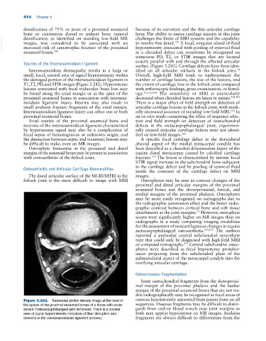
Figure 3.242. Transverse proton density image at the level of osseous hypointensity separated from parent bone on all
the apices of the proximal sesamoid bones of a horse with acute sequences. Osseous fragments may be difficult to distin
severe metacarpophalangeal joint lameness. There is a circular guish from end‐on blood vessels near joint margins as
area of signal hyperintensity indicative of fiber disruption and both may appear hypointense on MR images. Avulsion
desmitis in the intersesamoidean ligament (arrows). fragments are always difficult to differentiate from the