
Page 451 - Adams and Stashak's Lameness in Horses, 7th Edition
P. 451
Diagnostic Imaging 417
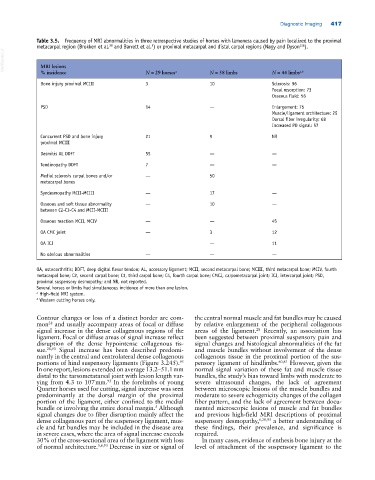
Table 3.5. Frequency of MRI abnormalities in three retrospective studies of horses with lameness caused by pain localized to the proximal
128
28
5
metacarpal region (Brokken et al. and Barrett et al. ) or proximal metacarpal and distal carpal regions (Nagy and Dyson ).
VetBooks.ir MRI lesions N = 29 horses a N = 58 limbs N = 44 limbs a,b
% incidence
Bone injury proximal MCIII 3 10 Sclerosis: 96
Focal resorption: 73
Osseous fluid: 56
PSD 14 — Enlargement: 75
Muscle/ligament architecture: 25
Dorsal fiber irregularity: 68
Increased PD signal: 57
Concurrent PSD and bone injury 21 9 NR
proximal MCIII
Desmitis AL DDFT 55 — —
Tendinopathy DDFT 7 — —
Medial sclerosis carpal bones and/or — 50
metacarpal bones
Syndesmopathy MCII‐MCIII — 17 —
Osseous and soft tissue abnormality — 10 —
between C2‐C3‐C4 and MCII‐MCIII
Osseous reaction MCII, MCIV — — 45
OA CMC joint — 3 12
OA ICJ — 11
No obvious abnormalities — — —
OA, osteoarthritis; DDFT, deep digital flexor tendon; AL, accessory ligament; MCII, second metacarpal bone; MCIII, third metacarpal bone; MCIV, fourth
metacarpal bone; C2, second carpal bone; C3, third carpal bone; C4, fourth carpal bone; CMCJ, carpometacarpal joint; ICJ, intercarpal joint; PSD,
proximal suspensory desmopathy; and NR, not reported.
Several horses or limbs had simultaneous incidence of more than one lesion.
a High‐field MRI system.
b Western cutting horses only.
Contour changes or loss of a distinct border are com the central normal muscle and fat bundles may be caused
mon and usually accompany areas of focal or diffuse by relative enlargement of the peripheral collagenous
28
28
signal increase in the dense collagenous regions of the areas of the ligament. Recently, an association has
ligament. Focal or diffuse areas of signal increase reflect been suggested between proximal suspensory pain and
disruption of the dense hypointense collagenous tis signal changes and histological abnormalities of the fat
sue. 28,93 Signal increase has been described predomi and muscle bundles without involvement of the dense
nantly in the central and centrolateral dense collagenous collagenous tissue in the proximal portion of the sus
portions of hind suspensory ligaments (Figure 3.245). pensory ligament of hindlimbs. 60,61 However, given the
93
In one report, lesions extended on average 13.2–51.1 mm normal signal variation of these fat and muscle tissue
distal to the tarsometatarsal joint with lesion length var bundles, the study’s bias toward limbs with moderate to
ying from 4.3 to 107 mm. In the forelimbs of young severe ultrasound changes, the lack of agreement
93
Quarter horses used for cutting, signal increase was seen between microscopic lesions of the muscle bundles and
predominantly at the dorsal margin of the proximal moderate to severe echogenicity changes of the collagen
portion of the ligament, either confined to the medial fiber pattern, and the lack of agreement between docu
bundle or involving the entire dorsal margin. Although mented microscopic lesions of muscle and fat bundles
5
signal changes due to fiber disruption mainly affect the and previous high‐field MRI descriptions of proximal
dense collagenous part of the suspensory ligament, mus suspensory desmopathy, 6,28,93 a better understanding of
cle and fat bundles may be included in the disease area these findings, their prevalence, and significance is
in severe cases, where the area of signal increase exceeds required.
30% of the cross‐sectional area of the ligament with loss In many cases, evidence of enthesis bone injury at the
of normal architecture. 5,6,93 Decrease in size or signal of level of attachment of the suspensory ligament to the