
Page 449 - Adams and Stashak's Lameness in Horses, 7th Edition
P. 449
Diagnostic Imaging 415
VetBooks.ir
A B
Figure 3.243. Sagittal and transverse proton density images of synovial fluid in the cartilage defect (arrows). A wavy, thin, hypoin
the fetlock of a horse with chronic metacarpophalangeal joint tense line overlying the cartilage defect may indicate the presence
lameness. There is an elliptical area of full‐thickness cartilage loss of pannus tissue along with synovial fluid (A). An irregular island of
on the dorsodistal aspect of the medial condyle of the third fluid hyperintensity associated with pooling of synovial fluid in the
metacarpal bone (arrows). This is characterized by replacement of chondral defect can be recognized on the transverse PD image that
the normal hypointense cartilage layer with pooling of hyperintense runs through the affected joint surface (B).
tendon or ligament in which they are embedded, due to
the similarities in signal intensity between bone, tendon,
and ligaments.
Abnormalities of the Digital Flexor Tendon Sheath
Injuries of the digital flexor tendons in the digital
flexor tendon sheath may be recognized as dispersed
small, focal areas of signal hyperintensity, distinct hyper
intense core lesions, thickening of the affected lobe(s),
and/or longitudinal parasagittal or frontal splits of the
lateral or medial border of the tendon with partial
separation of the tendon margins (Figure 3.244). Lesions
of the DDFT within the digital flexor tendon sheath
may continue distally into the navicular bursa and the
insertion on the distal phalanx. Lesions of the superfi
cial digital flexor tendon within the digital flexor tendon
sheath may extend into one of the branches of the ten
don and its insertion on the middle scutum. Areas of
signal hyperintensity and contour changes of the flexor
tendons within the digital sheath are most obvious in
transverse fat‐suppressed T1 spoiled gradient‐echo
(SPGR) images and frequently not visible ultrasono
graphically. Increased fluid distension and intrathecal
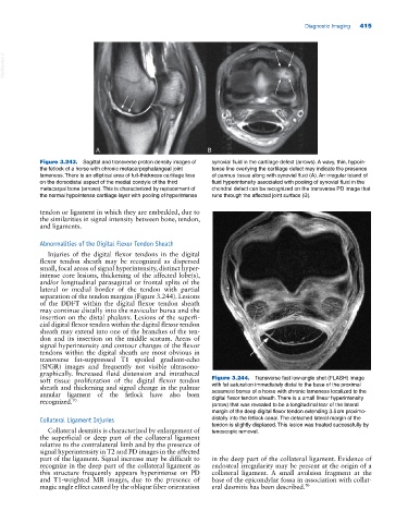
soft tissue proliferation of the digital flexor tendon Figure 3.244. Transverse fast low‐angle shot (FLASH) image
sheath and thickening and signal change in the palmar with fat saturation immediately distal to the base of the proximal
annular ligament of the fetlock have also been sesamoid bones of a horse with chronic lameness localized to the
recognized. 70 digital flexor tendon sheath. There is a small linear hyperintensity
(arrow) that was revealed to be a longitudinal tear of the lateral
margin of the deep digital flexor tendon extending 3.5 cm proximo
Collateral Ligament Injuries distally into the fetlock canal. The detached lateral margin of the
tendon is slightly displaced. This lesion was treated successfully by
Collateral desmitis is characterized by enlargement of tenoscopic removal.
the superficial or deep part of the collateral ligament
relative to the contralateral limb and by the presence of
signal hyperintensity in T2 and PD images in the affected
part of the ligament. Signal increase may be difficult to in the deep part of the collateral ligament. Evidence of
recognize in the deep part of the collateral ligament as endosteal irregularity may be present at the origin of a
this structure frequently appears hyperintense on PD collateral ligament. A small avulsion fragment at the
and T1‐weighted MR images, due to the presence of base of the epicondylar fossa in association with collat
magic angle effect caused by the oblique fiber orientation eral desmitis has been described. 70