
Page 446 - Adams and Stashak's Lameness in Horses, 7th Edition
P. 446
412 Chapter 3
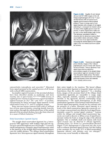
Figure 3.238. Sagittal (A) and dorsal
VetBooks.ir metacarpophalangeal joint of an 11‐year‐
(B) low‐field T2* GRE images of the left
old Warmblood with lameness due to
sagittal groove trauma of the proximal
phalanx. There is a large area of high
signal intensity with a margin of low signal
extending in a dorsopalmar direction along
the base of the sagittal groove (lower left
arrows). A similar signal abnormality can
be seen on the dorsal image (right arrow).
This fat/water cancelation artifact is
characteristic for abnormal osseous fluid
accumulation (bone contusion). Notice
also the area of low signal (sclerosis)
occupying the dorsal aspect of the sagittal
ridge of the third metacarpal bone (upper
A B left arrows).
Figure 3.239. Transverse and sagittal
proton density images of the metacar
pophalangeal joint of a horse with chronic
fetlock lameness. There is abnormal focal
signal hyperintensity within the
proximolateral aspect of the straight distal
sesamoidean ligament indicative of local
fiber disruption (black arrow). The lesion
originates at the distal border of the lateral
proximal sesamoid bone and extends
A B
11 mm distally (white arrow).
osteoarthritis (osteophytes and synovitis). Abnormal their entire length to the insertion. The lateral oblique
69
bone signal persisted at the sagittal groove of all horses distal sesamoidean ligament is frequently larger and more
with recheck MRI examinations. hyperintense than the medial ligament. In a standing
172
Incomplete sagittal fractures of the proximal phalanx horse, images of the proximal third of the oblique distal
have been described mainly in racehorses and more sesamoidean ligaments are susceptible to magic angle
recently also in sport horses. 58,69,91,99,144 MRI reviews of effect, especially medially, resulting in increased signal
sagittal groove trauma in sport horses report incomplete intensity in the ligaments, thereby confounding image
99
69
51
171
sagittal fractures in 20, 28, and 50% of patients, interpretation. The signal heterogeneity of the distal
characterized by linear increased signal intensity in the sesamoidean ligaments varies between individual horses.
172
subchondral bone in T1‐ and T2‐weighted images. Normal signal heterogeneity should not be confused with
Bone marrow lesions (osseous fluid) with or without the presence of a lesion. Abnormal signal intensity in a
loss of bone mineral density, extending distally from the lesion of the distal sesamoidean ligaments commonly cov
joint surface through the subchondral bone of the medial ers a larger cross‐sectional area than the focal signal hyper
172
glenoid of the proximal phalanx, have also been observed intensities caused by normal signal variation. Lesions
as a cause of fetlock lameness. 70,137,139,144 within the oblique and straight distal sesamoidean liga
ments can result in discrete or diffuse areas of signal hyper
intensity within the body or along the edge of the affected
Distal Sesamoidean Ligament Injuries
ligament. One or multiple small core lesions with focal
The straight distal sesamoidean ligament has a hetero signal increase may be observed in affected ligaments,
geneous MRI appearance with multiple high and low sig extending from 5 to 30 mm in a proximodistal direction
nal areas spread throughout most of its length. A normal, (Figure 3.239). Enlargement of a ligament may also occur
172
small homogeneous triangular signal hyperintensity exists with or without abnormal signal increase, but this finding
at the insertion of the straight distal sesamoidean ligament is less consistent. In one study, 30% of distal sesamoidean
70
onto the middle phalanx. The oblique distal sesamoidean ligaments affected with desmitis were not enlarged.
ligaments have heterogeneous signal intensity throughout Lesions may occur biaxially or bilaterally.