
Page 444 - Adams and Stashak's Lameness in Horses, 7th Edition
P. 444
410 Chapter 3
had subchondral bone abnormalities of the distal meta in less than 10% of racehorses with fetlock lameness,
144
carpus and one third lesions of the distal sesamoidean although another more recent study found evidence of
VetBooks.ir horses, also from a variety of breeds and disciplines, sub Thoroughbreds and 37% of racing Standardbreds, but
periarticular soft tissue injuries in 21% of racing
ligaments. However, in a different MRI study of 232
70
these were rarely considered the main cause of lameness
chondral bone abnormalities were very uncommon,
while more than half the horses were lame because of (Table 3.4). 139
distal sesamoidean ligament injuries. On the other
90
hand, an MRI study of 131 racing Thoroughbreds with
fetlock lameness reported almost exclusively osseous MRI Abnormalities in the Fetlock Region
injuries, including subchondral bone trauma and incom Subchondral Bone Abnormalities of the Metacarpal/
plete stress fractures. In both mixed breed studies, 70,90
144
distal sesamoidean ligament desmitis was the most com Metatarsal Condyles
mon soft tissue injury, while other soft tissue injuries of The normal subchondral bone thickness of the distal
the suspensory branches, collateral ligaments, intersesa aspect of the MCIII/MTIII varies from dorsal to palmar
moidean ligaments, and flexor tendons also occurred but and from abaxial to axial, being thinnest axially and
with much lower frequency. Cartilage lesions, osteoar thickest in the middle of each condyle, especially
thritis, and osteochondral lesions were also reported. toward the palmar aspect. Subchondral bone thick
47
Combinations of injuries were common in the fetlock ness of the distal aspect of the MCIII/MTIII is likely to
region, with 63% of horses in one study being identified change as it adapts to the type of exercise the horse
70
with multiple MRI abnormalities. Most common was performs. The subchondral bone thickness of the prox
the combination between subchondral bone lesions and imal phalanx increases slightly toward the palmar
injuries involving components of the suspensory appara aspect of each condyle. There is reasonable laterome
tus. Abnormalities of the subchondral bone and lesions dial symmetry in subchondral bone thickness of both
70
of the distal sesamoidean ligaments have also been docu the MCIII/MTIII and the proximal phalanx. The
mented in other MRI reports. 54,151,172 Although subchon increased use of MRI has highlighted the importance
dral bone injuries have been diagnosed with standing and high prevalence of bone sclerosis and bone marrow
low‐field MRI, soft tissue abnormalities are much less lesions in subchondral bone injury in the fetlock of
166
144
commonly identified using standing low‐field MRI. horses. 42,51,69,139
The most common MRI diagnosis in racing Abnormal MRI signal in POD lesions of the condyles
Thoroughbreds was palmar/plantar osteochondral dis of the MCIII/MTIII is manifested as diffuse or focal
ease (POD) of the MCIII/MTIII, present in more than signal increase in fat‐suppressed images consistent with
half of the horses examined. 144 Fractures were also bone marrow lesion or bruising and diffuse T1, PD, and
much more commonly diagnosed on MR images of the T2 signal decrease consistent with trabecular thickening
fetlock of racing Thoroughbreds compared with other and osteosclerosis (Figure 3.236). Osseous fluid appears
disciplines. 140,141,144,183 Soft tissue injuries were identified more commonly in the acute stage of injury, while
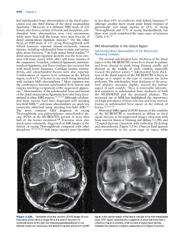
A B
Figure 3.236. Transverse short tau recovery (STIR) image (A) and signal in the palmar aspect of the lateral condyle of the third metacarpal
transverse proton density image (B) at the level of the proximal bone. STIR signal hyperintensity suggestive of abnormal bone fluid is
sesamoid bones of the right fore metacarpophalangeal joint of two present in Figure A (arrows), while an area of signal hypointensity
different 3‐year‐old racehorses with fetlock lameness and abnormal MR indicates the presence of palmar osteosclerosis in Figure B (arrows).