
Page 439 - Adams and Stashak's Lameness in Horses, 7th Edition
P. 439
Diagnostic Imaging 405
VetBooks.ir
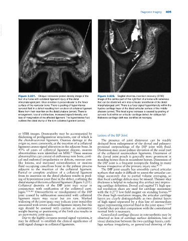
Figure 3.231. Oblique transverse proton density image of the Figure 3.232. Sagittal short tau inversion recovery (STIR)
foot of a horse with collateral ligament injury of the distal image of the central part of the right foot of a horse with lameness
interphalangeal joint. Slice direction is perpendicular to the flexor that can be abolished with intra‐articular anesthesia of the distal
surface of the navicular bone. There is pooling of hyperintense interphalangeal joint. There is a focal signal hyperintensity within the
synovial fluid in a defect resulting from avulsion of collateral ligament hyaline cartilage layer of the distal articular surface of the middle
fibers from their insertion on the distal phalanx (arrow). There is phalanx (arrow). This focal signal increase is caused by pooling of
enlargement, loss of architecture, increased signal intensity, and synovial fluid within an articular cartilage defect. An oblique full‐
loss of margination of the affected ligament. The hyperintense fluid thickness cartilage cleft was identified at necropsy.
outlines the distal stump of the torn collateral ligament (arrow).
or STIR images. Desmopathy may be accompanied by
thickening of periligamentar structures, one of which is Lesions of the DIP Joint
the chondrocoronal ligament. Osseous damage at the The presence of joint distension can be readily
origin or, more commonly, at the insertion of a collateral deduced from enlargement of the dorsal and palmaro
ligament causes signal alteration in the adjacent bone. In proximal outpouchings of the DIP joint with fluid.
45% of cases of collateral ligament disease, osseous Distension may cause palmar deviation of the axial part
39
abnormalities were identified on MRI. These osseous of the collateral sesamoidean ligaments. Distension of
abnormalities can consist of entheseous new bone, corti the dorsal joint pouch is generally more prominent in
cal and endosteal irregularities or defects, osseous cyst‐ standing horses than in recumbent horses. Distension of
like lesions, and increased mineralization or osseous the DIP joint is a frequent nonspecific finding in many
fluid occupying cancellous bone in the palmar process horses irrespective of the primary injury site. 46,152
adjacent to the insertion of the collateral ligament. The DIP joint usually has smoothly curved articular
Partial or complete avulsion of a collateral ligament surfaces that make it difficult to assess the articular car
from its insertion on the distal phalanx results in pool tilage accurately due to partial volume averaging, so
ing of hyperintense joint fluid in the defect, outlining the that focal cartilage defects may be missed. Thinner slice
distal stump of the torn collateral ligament (Figure 3.231). thickness is helpful in reducing this artifact and improv
Collateral desmitis of the DIP joint may occur in ing cartilage definition. Dorsal and sagittal T1 high spa
conjunction with ossification of the collateral carti tial resolution slices are used for cartilage assessment
lages. 55,56,105 Osteoarthritis or misalignment of the DIP with the 0.27 T low‐field magnet on standing horses.
149
joint caused by subluxation associated with collateral On these high‐resolution images, the articular cartilage
ligament injury may occur but are rare. Marked layers are clearly distinguishable as two distinct layers
widening of the joint space may indicate joint instability of high signal separated by a thin line of intermediate
associated with severe collateral ligament injury, but this signal representing synovial fluid in the joint space. 133,149
sign should be assessed with caution as asymmetric Careful slice‐per‐slice comparison with the contralateral
weight‐bearing or positioning of the limb also results in normal limb is necessary.
an asymmetric joint space. Generalized cartilage disease in osteoarthritis may be
Due to the highly common normal signal variation, it observed as loss of cartilage surface definition, loss of
may be difficult to establish the clinical significance of visual distinction between the two cartilage layers, carti
mild signal changes in collateral ligaments. lage surface irregularity, or generalized thinning of the