
Page 438 - Adams and Stashak's Lameness in Horses, 7th Edition
P. 438
404 Chapter 3
VetBooks.ir
A B
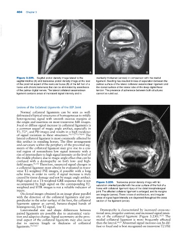
Figure 3.229. Sagittal proton density image lateral to the markedly thickened (arrows) in comparison with the medial
sagittal midline (A) and transverse proton density image at the level ligament. Swelling has resulted in loss of separation between the
of the proximal aspect of the navicular bursa (B) of the left foot of a palmar surface of the lateral collateral sesamoidean ligament and
horse with chronic lameness that can be eliminated by anesthesia the dorsal surface of the lateral lobe of the deep digital flexor
of the palmar digital nerves. The lateral collateral sesamoidean tendon. The presence of adherence between both structures
ligament contains areas of increased signal intensity and is cannot be ruled out.
Lesions of the Collateral Ligaments of the DIP Joint
Normal collateral ligaments can be seen as well
delineated elliptical structures of homogeneous to mildly
heterogeneous signal with smooth osseous margins at
the origin and insertion on most transverse MR images.
Focal or diffuse signal increase in collateral ligaments is
a common sequel of magic angle artifact, especially in
T1, T2*, and PD images and results in a high incidence
of signal variation in these structures. 74,171,177,178,192 The
lateral collateral ligament is most commonly affected by
this artifact in standing horses. The fiber arrangement
and curvature within the periphery of the proximal seg
ments of the collateral ligament may give rise to a cen
tral region of nonuniform low signal intensity with a
rim of intermediate to high signal intensity at the level of
the middle phalanx due to magic angle effect that can be
confused with a desmopathy on both low‐ and high‐
field images. 90,192 Therefore, suspected signal changes in
a collateral ligament must always be evaluated in trans
verse T2‐weighted FSE images, if possible with a long
echo time, in order to verify if signal increase is truly
caused by tissue damage and not by magic angle artifact.
High signal on a T1‐weighted GRE sequence that is not
accompanied by high signal on the corresponding T2‐ Figure 3.230. Transverse proton density image with fat
weighted and STIR images is not a reliable indicator of saturation oriented parallel with the solar surface of the foot of a
horse with collateral ligament injury of the distal interphalangeal
injury. joint. The affected collateral ligament is enlarged, and its margins
On dorsal images obtained in an image plane parallel are irregular (arrow). There is loss of architecture, and irregular
with the direction of the collateral ligaments and per areas of signal hyperintensity are dispersed throughout the cross
pendicular to the solar surface of the foot, the collateral section of the ligament (arrow).
ligaments appear as curved, banana‐shaped bands of
homogeneous, low T2 signal.
Lateromedial size and shape differences between Desmopathy is characterized by increased cross‐sec
paired ligaments are possible due to anatomical varia tional area, irregular contour, and increased signal inten
tion and adaptive change. Signal asymmetry at the prox sity of the collateral ligament (Figure 3.230). 44,73 The
imal aspect of the collateral ligaments may also occur medial collateral ligament is more frequently affected
due to uneven length or thickness of collateral than the lateral. 44,53 Abnormal signal increase can be dif
ligaments. 44,53,117 fuse or focal and is best recognized on transverse T2 FSE