
Page 447 - Adams and Stashak's Lameness in Horses, 7th Edition
P. 447
Diagnostic Imaging 413
The location of lesions within distal sesamoidean
ligaments and the distribution of lesions varies between
VetBooks.ir more commonly in the distal part of the ligament, proxi
Straight sesamoidean desmitis occurs
studies.
70,90,151,172
although
mal to its insertion on the middle phalanx,
151,172
proximal lesions near the origin were most common in
another study (Figure 3.239). Oblique distal sesamoid
70
ean desmitis can occur proximally or throughout the
entire length of the ligament. 70,90,151,172 Cruciate distal
sesamoidean desmitis is very rare. 144,172
Reports suggest that distal sesamoidean desmitis is fre
quently regarded as the primary cause of lameness, 70,90,151
although one author considered lesions to be the sole
cause of lameness in only 2 of 58 horses with evidence of
desmitis. The majority of horses with oblique or
172
straight distal sesamoidean desmitis diagnosed with MRI
do not have a palpable enlargement, nor do they show
any ultrasonographic abnormalities. 70,151,172
Suspensory Ligament Branch Injuries
The suspensory ligament branches are paired triangu
lar structures of low signal intensity that flatten in a dor
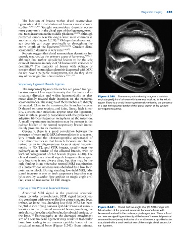
sopalmar direction and widen lateromedially as they Figure 3.240. Transverse proton density image of a metatar
move distally toward their insertion on the proximal sophalangeal joint of a horse with lameness localized to the fetlock
sesamoid bones. The margins of the branches are sharply region. There is a small, linear hyperintensity reflecting the presence
delineated. Close to the insertion, the branches become of a tear in the plantar border of the lateral branch of the suspen
D shaped on cross section, and faint, linear, high inten sory ligament (arrow).
sity, dorsopalmar striations appear near the ligament–
bone interface, possibly associated with the presence of
adaptive fibrocartilaginous metaplasia at the insertion.
A small hyperintense indentation may be present in the
palmar border of the normal suspensory branch imme
diately proximal to its insertion.
Generally, there is a good correlation between the
presence of (even mild) MRI abnormalities in a suspen
sory branch and the ultrasonographic appearance of
fiber abnormalities in that branch. Lesions are charac
terized by an intraligamentous focus of signal hyperin
tensity in PD, T2, and STIR images, usually near the
palmar/plantar border of the affected branch, with or
without enlargement of that branch (Figure 3.240). The
clinical significance of mild signal changes in the suspen
sory branches is not always clear, but they may be the
only finding in an otherwise normal MRI examination
of a horse whose lameness was abolished by a low four‐
point nerve block. During standing low‐field MRI, false
signal increase in one or both suspensory branches may
be caused by vascular flow artifact or magic angle arti
fact, even on transverse T2 FSE images.
Injuries of the Proximal Sesamoid Bones
Abnormal MRI signal in the proximal sesamoid
bones includes osteosclerosis, STIR signal hyperinten
sity consistent with osseous fluid or contusion, and focal
trabecular bone loss. Standing low‐field MRI has been
helpful in identifying osseous cyst‐like lesions at various Figure 3.241. Dorsal fast low‐angle shot (FLASH) image with
locations in the proximal sesamoid bones, involving the fat saturation of the proximal sesamoid bones of a horse with
articular surface, the axial border, the abaxial surface, or lameness localized to the metacarpophalangeal joint. There is focal
the base. Enthesopathy at the damaged attachment and intense signal hyperintensity at the base of the medial proximal
108
site of a sesamoidean ligament may result in trabecular sesamoid bone (arrow) indicative of a small osseous cyst‐like lesion
bone loss leading to an osseous cyst‐like lesion of the associated with a small vertical tear of the straight distal sesamoid
proximal sesamoid bone (Figure 3.241). Bone mineral ean ligament.