
Page 452 - Adams and Stashak's Lameness in Horses, 7th Edition
P. 452
418 Chapter 3
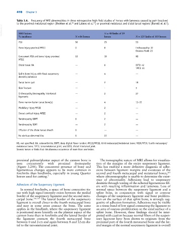
Table 3.6. Frequency of MRI abnormalities in three retrospective high‐field studies of horses with lameness caused by pain localized
94
28
6
to the proximal metatarsal region (Brokken et al. and Labens et al. ) or proximal metatarsal and distal tarsal regions (Barrett et al. ).
VetBooks.ir MRI lesions N = 16 horses N = 48 limbs of 39 N = 125 limbs of 103 horses
% incidence
horses
PSD 50 25 53
Bone injury proximal MTIII 0 8 Enthesopathy: 32
Osseous fluid: 23
Concurrent PSD and bone injury proximal 10 19 NS
MTIII
Distal tarsal OA — 6 DITJ: 42
TMTJ: 51
Splint bone injury with focal suspensory — 4 —
desmitis/adhesion
Tarsal bone cyst — 2 18
Slab fracture — ‐ 3
Enthesopathy/desmopathy intertarsal — 2 27
ligaments
Bone marrow lesion tarsal bone(s) — 4 13–22
Medullary injury MTIII — 2 —
Dorsal cortical injury MTIII — 2 —
Tendinopathy DDFT 6 2 —
Tendinopathy SDFT — 2 —
Effusion of the distal tarsal sheath 6 — —
No obvious abnormalities 6 22 —
NS, not specified; OA, osteoarthritis; DDFT, deep digital flexor tendon; MCIII/MTIII, third metacarpal/metatarsal bone; MCIV/MTIV, fourth metacarpal/
metatarsal bone; TMTJ, tarsometatarsal joint; and DITJ, distal intertarsal joint.
Several horses or limbs had simultaneous occurrence of more than one lesion.
proximal palmar/plantar aspect of the cannon bone is The tomographic nature of MRI allows for visualiza
seen concurrently with proximal desmopathy tion of the margins of the entire suspensory ligament.
(Figure 3.246). The concurrent presence of bony and This has enabled a more definitive diagnosis of adhe
ligamentous changes appears to be more common in sions between ligament margins and exostoses of the
203
forelimbs than hindlimbs, especially in young Quarter second and fourth metacarpal and metatarsal bones,
horses used for cutting. 5 where ultrasonography is unable to determine the exist
ence of abnormality. Adhesions lead to suspensory
desmitis through tearing of the adhered ligamentous fib
Adhesions of the Suspensory Ligament
ers with resulting inflammation and lameness. Loss of
In normal forelimbs, a space of loose connective tis normal space between the suspensory ligament and a
sue with high signal intensity exists between the medial splint bone, in conjunction with signal or contour
border of the suspensory ligament and the second meta changes of the suspensory ligament and bone prolifera
carpal bone. 124,127 The lateral border of the suspensory tion on the surface of that splint bone, is strongly sug
ligament is overall closer to the fourth metacarpal bone gestive of adhesion formation. Adhesions may be visible
and may in some areas contact the bone. The same as a tissue band of low signal connecting the ligament to
applies in the hindlimb, where the suspensory ligament an area of osseous proliferation on the axial surface of a
is positioned more laterally on the plantar aspect of the splint bone. However, these findings should be inter
cannon bone than in forelimbs and the lateral border of preted with caution because normal fibers of the suspen
the ligament contacts the fourth metacarpal bone sory ligament have been shown to originate from the
between 0 and 2 cm and again between 8 and 12 cm dis proximal part of the fourth metatarsal bone and the lat
tal to the tarsometatarsal joint. eral margin of the normal suspensory ligament is overall