
Page 457 - Adams and Stashak's Lameness in Horses, 7th Edition
P. 457
Diagnostic Imaging 423
fractures of the talus or calcaneus that are not readily also occur in the fourth tarsal bone or in the distal tibia
visible radiographically. 33 at the apex of the parasagittal grooves that articulate
VetBooks.ir Osseous Cyst‐like Lesions lesions have intermediate to high signal intensity sur
with the trochlear ridges of the talus. Osseous cyst‐like
rounded by low signal intensity of sclerotic bone. They
Osseous cyst‐like lesions occurred in 22% of limbs in may be incidental findings in the tarsal bones, especially
one MRI study of horses with distal tarsal region lame if there are no signal alterations in the surrounding
ness and were approximately equally distributed bone. Reactive signal changes that represent more sig
between the third and central tarsal bones. They may nificant bone damage are osseous fluid or extensive scle
6
rosis in surrounding bone. Osseous cyst‐like lesions are
usually associated with an articular surface defect.
Intertarsal Ligament Injury
Lesions of the intertarsal interosseous ligament
occurred in 33% of horses with distal tarsal region
lameness. Lesions have been described as either signal
6
increase or signal decrease in the ligament with loss of
6
57
normal ligament architecture. Changes of the ligament
fossa include thickening and irregular contour of the
subchondral bone and narrowing to even total oblitera
tion of the fossa by new bone production. Osseous fluid
and/or sclerosis may be present in the third and central
tarsal bones either side of the ligament fossa
(Figure 3.249). Abnormalities of the ligament and the
ligament fossa typically occur concurrently. Severe
changes of desmopathy and enthesopathy are invariably
accompanied by degenerative changes of the distal inter
tarsal joint. 6
Other Soft Tissue Injuries of the Tarsus
Care is required when interpreting signal increase
because several ligaments in the tarsus are subject to
Figure 3.248. Transverse proton density image through the magic angle artifact, both in standing and recumbent
proximal row of tarsal bones of the left tarsus of a horse with focal MRI, which can overlap with certain types of injury.
12
osteoarthritis of the articulation between the central and fourth
tarsal bones. There is loss of joint space and subchondral bone Anecdotal reports have illustrated medial and lateral
167
outline surrounded by a margin of irregular osteosclerosis of the collateral ligament injuries. Hyperintense core lesions
fourth and central tarsal bones (arrow). of the long plantar ligament may also occur, and
A B
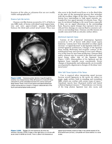
Figure 3.249. Sagittal (A) and transverse (B) short tau signal hyperintensity (osseous fluid) in the plantar aspect of the
inversion recovery (STIR) images of the right tarsus of a horse with third tarsal bone (arrows), compatible with bone edema or a bone
acute onset hindlimb lameness. There is abnormal intraosseous bruise.