
Page 456 - Adams and Stashak's Lameness in Horses, 7th Edition
P. 456
422 Chapter 3
sequences, but the dorsal half is more heterogeneous
with multifocal spots or lines or bands of higher signal
VetBooks.ir frequently performed together, as diagnostic anesthesia
intensity.
59
MRI of the tarsal and proximal metatarsal regions is
is insufficiently specific for accurate localization of pain
causing lameness in this area. 6,93 In one study, 47% of
horses with a positive response to subtarsal perineural
diagnostic anesthesia were found to have a primary
lesion in the distal tarsal joints, while 33% of horses
with a positive response to intra‐articular anesthesia of
the distal tarsal joints were diagnosed with proximal
suspensory desmitis. Therefore, MRI protocols should
6
be adopted to allow for complete evaluation of both
anatomic regions in horses where pain has been local
ized to this area and conventional imaging techniques
produce negative or equivocal results.
Several clinical reports and one large case series of
in vivo tarsal MRI have become available in recent
years. 6,13,23,33,40,41,57,93,96,131,167,169 The prevalence of MRI
lesions of the distal tarsus detected with a high‐field
magnet was reported for 125 limbs of 103 horses with
lameness originating from the distal tarsal/proximal
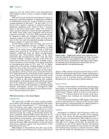
metatarsal area. Osteoarthritic changes of the distal Figure 3.247. Sagittal proton density image of the tarsus of a
intertarsal and tarsometatarsal joints were the most horse with osteoarthritis of the talocalcaneal joint. There is localized
common findings, including sclerosis of the third and loss of joint space and subchondral bone margins in the center of
central tarsal bones in 54% and 100% of limbs, respec the joint. This lesion is surrounded by a wide irregular area of signal
tively, osteophytes at the margins of the distal intertarsal loss reflecting reactive osteosclerosis in both the talus and the
or tarsometatarsal joints in 51% and 70% of limbs, sustentaculum tali of the calcaneus (arrows).
respectively, and articular cartilage damage or subchon
dral bone lysis of the same joints in 42% and 51% of
limbs, respectively. Degenerative changes of osteoarthri
tis (osteolysis and loss of joint space) were also found in (Figure 3.248) and in the talocalcaneal joint (Figure 3.247).
65% limbs between the central and fourth, central and However, mild subchondral bone contour irregularities,
second, third and second, and third and fourth tarsal sclerosis, osteophytes, and intertarsal ligament changes
bones. Suspensory ligament desmopathy was found in may be seen in non‐lame horses, and their presence
53% of limbs in this study, suggesting that multiple should not be overinterpreted as clinical disease.
abnormalities of the distal tarsal joints and the proximal
part of the suspensory ligament were frequently identi Osseous Trauma
fied simultaneously in this population of predominantly
Western performance horses. No retrospective MRI Osseous trauma related to trabecular microfracture,
studies are available to document the prevalence of inju bone edema, or hemorrhage characterized by high STIR
ries of the proximal tarsal region. signal in bone can occur in the central, third, and fourth
tarsal bones 6,13 (Figure 3.249) as well as in the talus or
calcaneus. 93,202 Osseous fluid in the vicinity of the inter
MRI Abnormalities in the Tarsal Region tarsal foramen may represent an intertarsal ligament
enthesopathy.
Osteoarthritis The most common finding on MR images of the tarsus
The superiority of MRI over other imaging modali of horses with tarsal pain is sclerosis of the central and/or
ties to detect early changes of osteoarthritis in the tarsus third tarsal bones, characterized by low signal intensity in
has been clearly demonstrated. 6,26,40,96 Periarticular new the medulla and/or subchondral bone region. 6,40
bone formation, joint space narrowing, subchondral
sclerosis, and subchondral bone lysis may be seen in the Fractures
tarsometatarsal, distal intertarsal, and rarely proximal
intertarsal joints. In a study of 38 Icelandic horses, the Incomplete or complete vertical fractures of the cen
most commonly detected MRI lesions observed in 42 tral (or third) tarsal bone are usually stress fractures
distal intertarsal joints classified as osteoarthritic were and may not always be detected radiographically. 6,40
mineralization front defects, joint margin lesions, and These fractures are identified on MR images as a line of
articular cartilage lesions. An association has been high signal intensity in most imaging sequences but
96
reported between MRI abnormalities of the intertarsal especially on T1 images, where they are generally sur
interosseous ligament region and osteoarthritis of the rounded by an area of low signal intensity representing
distal intertarsal joint. 6,169 osseous fluid surrounding an acute fracture or sclerosis
6
MRI changes of osteoarthritis may also occur in the surrounding stress fractures or chronic fractures. MRI
vertical contact areas between the various tarsal bones may also be useful for identification of non‐displaced