
Page 891 - Adams and Stashak's Lameness in Horses, 7th Edition
P. 891
Principles of Musculoskeletal Disease 857
A more recent diagnostic tool called sonoelastogra
phy is currently being investigated for diagnostic use for
VetBooks.ir stiffness of tissues and may be able to distinguish
tendonopathy.
This technology can assess the inner
38,39
between different phases (acute, subacute, and chronic)
of healing with much more accuracy than traditional
ultrasound. If further investigation confirms initial
promising data, this monitoring technique will be a
mainstay in monitoring tendon response to rehabilita
tion and prognostic outcomes.
BIOMARKERS FOR TENDON DISEASE
The field of biomarkers is of keen interest to many
clinicians and researchers. Accurate detection of sub
clinical disease contributes to improved prevention of
a common career‐ending disease. Furthermore, early
detection of disease enables more tailored rehabilitation
programs and altered training regimens. Ultrasound
remains the gold standard of detection, permitting a
16
more accurate reflection of cellular disease and sensitiv
ity to subtle changes in the tissue state. Biomarkers that
detect the stage of disease may also predict optimal
treatment and rehabilitation protocols and evaluate
their efficacy.
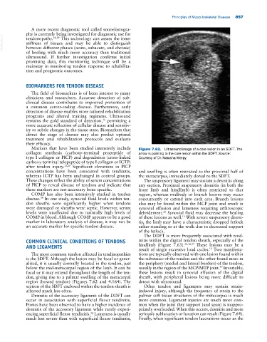
Markers that have been studied intensively include Figure 7.62. Ultrasound image of a core lesion in an SDFT. The
collagen synthesis (carboxy‐terminal propeptide of arrow is pointing to the core lesion within the SDFT. Source:
type I collagen or PICP) and degradation (cross‐linked Courtesy of Dr. Natasha Werpy.
carboxy‐terminal telopeptide of type I collagen or ICTP)
after tendon injury. 12,20 Significant elevations in PICP
concentrations have been associated with tendinitis, and swelling is often restricted to the proximal half of
whereas ICTP has been unchanged in control groups. the metacarpus, immediately dorsal to the SDFT.
These changes reflect the ability of serum concentrations The suspensory ligament may sustain a desmitis along
of PICP to reveal disease of tendons and indicate that any section. Proximal suspensory desmitis (in both the
these markers are not necessary bone specific. front limb and hindlimb) is often restricted to that
COMP has also been intensively studied in tendon region, whereas midbody or branch lesions may occur
disease. In one study, synovial fluid levels within ten concurrently or extend into each area. Branch lesions
34
don sheaths were significantly higher when tendons also may be found within the MCP joint and result in
were damaged or sheaths were septic. However, serum synovial effusion and lameness requiring arthroscopic
levels were unaffected due to naturally high levels of debridement. Synovial fluid may decrease the healing
26
COMP in blood. Although COMP appears to be a good of these lesions as well. With severe suspensory desmi
12
marker in laboratory analysis of disease, it may not be tis, the limb may have a characteristic dropped fetlock
an accurate marker for specific tendon disease. either standing or at the walk due to decreased support
of the fetlock.
The DDFT is more frequently associated with tend
COMMON CLINICAL CONDITIONS OF TENDONS initis within the digital tendon sheath, especially of the
36,42,47
AND LIGAMENTS hindlimb (Figure 7.63). These lesions may be a
result of single excessive load cycles. Two manifesta
12
The most common tendon affected in tendinopathies tions are typically observed with one lesion found within
is the SDFT. Although the lesion may be focal or gener the substance of the tendon and the other found more in
alized, it is usually centrally located in the tendon, just the periphery (medial and lateral borders) of the tendon,
47
below the mid‐metacarpal region of the limb. It can be usually in the region of the MCP/MTP joint. Invariably,
focal or it may extend throughout the length of the ten these lesions result in synovial effusion of the digital
don, giving rise to a palmar swelling of the metacarpal sheath, with peripheral lesions being more difficult to
region (bowed tendon) (Figures 7.62 and 4.164). The detect with ultrasound.
section of the SDFT enclosed within the tendon sheath is Other tendon and ligaments may sustain strain‐
affected much less often. induced injury, although the frequency of strain to the
Desmitis of the accessory ligament of the DDFT can palmar soft tissue structures of the metacarpus is much
occur in association with superficial flexor tendinitis. more common. Ligament injuries are much more com
Ponies have been observed to have a higher incidence of mon when the joint they support (and span) is inappro
desmitis of the accessory ligament while rarely experi priately overloaded. When this occurs, desmitis and more
12
encing superficial flexor tendinitis. Lameness is usually seriously subluxation or luxation can result (Figure 7.64).
much less severe than with superficial flexor tendinitis, Finally, when significant tendon lacerations occur as the