
Page 889 - Adams and Stashak's Lameness in Horses, 7th Edition
P. 889
Principles of Musculoskeletal Disease 855
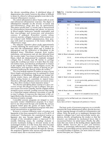
the chronic remodeling phase. A subclinical phase of Table 7.1. A standard exercise program recommended following
degeneration also exists; however, this phase is difficult tendon injury.
VetBooks.ir minimal inflammatory reaction. Exercise
to detect by clinical or ultrasonographic exam due to the
The initial inflammatory phase begins with an acute
and substantial inflammatory response. The degree of level Week Duration and nature of exercise
inflammation depends on the severity of lesion and
anti‐inflammatory drugs that may be administered. 0 0–2 Box rest
This phase usually lasts 1–2 weeks and is characterized 1 3 10‐min walking daily
by intratendinous hemorrhage and edema, an increase
in blood supply, leukocytes (initially neutrophils and 1 4 15‐min walking daily
then macrophages and monocytes), and proteolytic
enzymes if unabated. Proteolytic enzyme release 1 5 20‐min walking daily
removes necrotic collagen; however, if the enzymes are
not reduced with NSAID administration, their pres 1 6 25‐min walking daily
ence will result in expansion of the lesion within the 1 7 30‐min walking daily
inflammatory stage.
The subacute reparative phase peaks approximately 1 8 35‐min walking daily
3 weeks following the initial injury. This phase over
32
laps with the inflammatory phase and is marked by 1 9 40‐min walking daily
angiogenesis and the infiltration of fibroblasts in the 1 10–12 45‐min walking daily
damaged tissue. Fibroblasts originate from tendon,
endotenon, and paratenon and are also delivered from Week 12: Repeat ultrasound examination
7
the vascular origin. Scar tissue is formed from the fibro
blasts, which are characterized by randomly arranged 2 13–16 40‐min walking and 5‐min trotting daily
collagen that is initially type III. Similar to cartilage
repair, the resulting scar tissue is weaker than the origi 2 17–20 35‐min walking and 10‐min trotting daily
nal tendon tissue, and therefore predisposed to reinjury 2 21–24 30‐min walking and 15‐min trotting daily
at the original site of injury. When reinjury occurs, the
inflammatory and reparative phases of healing are per Week 24: Repeat ultrasound examination
petuated, furthering the damage within the tendon. 32
The poor healing response of tendon in areas of poor 3 25–28 25‐min walking and 20‐min trotting daily
blood supply and paratenon may be explained by a lack 3 29–32 20‐min walking and 25‐min trotting daily
of migration of fibroblasts. Adhesions are formed fol
lowing tendon injury, and although detrimental, they Week 32: Repeat ultrasound examination
allow healing factors to contribute to injured tissue.
Therefore, although adhesions restrict movement of ten 4 33–40 45‐min exercise daily, gradually increasing
don, they allow tendon to form reparative tissue. 32 in amount
The chronic remodeling phase consists of a slow con
version of type III collagen to type I collagen. This pro 4 41–48 45‐min exercise daily with fast work 3
times a week
cess occurs over several months, and the original tendon
strength is never restored. Loading (exercise) that ensues Week 48: Repeat ultrasound examination
in a controlled manner enhances conversion of type III
to type I collagen. It improves the alignment of the col 5 48+ Return to full competition/race training
lagen fibrils in the direction of force and results in better
mechanical tendon strength. This protocol can be modified based upon ultrasound recheck
Controlled exercise programs are integral to every examinations.
successful tendon rehabilitation program. It is impor Source: Davis and Smith. Reproduced with permission of Elsevier.
12
tant to use ultrasound therapy and modify the exercise
program based upon the quality of repair. Table 7.1
illustrates a standard rehabilitation program with ultra The basic goals of treatment programs are to initially
sound reexamination at specified times. minimize the acute inflammatory phase, thereby decreas
Reinjury is common following repair of tendons and ing the ongoing damage of tendinous inflammation and
ligaments; it has been reported to occur in 8%–43% of edema. Next, the goals are to implement therapies that
racehorses. Furthermore, the contralateral tendon or improve collagen type I and extracellular matrix pro
14
ligament also may become injured. Although the duction, thereby strengthening the tendon so that the
14
strength of the remodeled tendon is improved 15–18 structure can withstand the mechanical forces that it
months following injury, the resulting elasticity is encountered before the initial injury. Finally, the adhe
severely weakened, leading to elevated strains in the sions that may form in the healing phases significantly
undamaged tendon. It is this tissue that often becomes reduce athletic function due to pain and lameness.
damaged when reinjury occurs. Furthermore, if the Therefore, minimizing inflammation, enhancing regen
SDFT is damaged and lengthened through the damage, eration, and controlling exercise optimize the chances of
the suspensory ligament may sustain microdamage and successful healing of tendons. For more detail on intral
eventual injury due to increased strains. 22 esional therapies, see Chapter 8.