
Page 885 - Adams and Stashak's Lameness in Horses, 7th Edition
P. 885
Principles of Musculoskeletal Disease 851
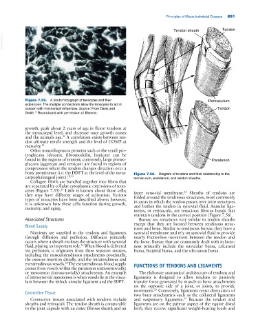
Tendon sheath Tendon
VetBooks.ir
Figure 7.55. A photomicrograph of tenocytes and their Retinaculum
extensions. The multiple connections allow the tenocytes to act in
concert with mechanical influences. Source: From Davis and Tendon
12
Smith. Reproduced with permission of Elsevier.
growth, peak about 2 years of age in flexor tendons at
the metacarpal level, and decrease once growth ceases
and the animals age. A correlation exists between ten
35
don ultimate tensile strength and the level of COMP at
maturity. 34
Other noncollagenous proteins such as the small pro
teoglycans (decorin, fibromodulin, lumican) can be
found in the regions of tension; conversely, large proteo Paratenon
glycans (aggrecan and versican) are found in regions of
compression where the tendon changes direction over a
bony prominence (i.e. the DDFT at the level of the meta Figure 7.56. Diagram of tendons and their relationship to the
carpophalangeal joint). 12,13 retinaculum, endotenon, and tendon sheaths.
Collagen fibrils are bundled together into fibers that
are separated by cellular cytoplasmic extensions of teno
cytes (Figure 7.55). Little is known about these cells; 24
25
they may have different stages of activation. Various inner synovial membrane. Sheaths of tendons are
folded around the tendinous structures, most commonly
types of tenocytes have been described above; however,
it is unknown how these cells function during growth, in areas in which the tendon passes over joint structures
and bathes the tendon in synovial fluid. Annular liga
maturity, and aging.
ments, or retinacula, are tenacious fibrous bands that
maintain tendons in the correct position (Figure 7.56).
Associated Structures Bursae are structures very similar to tendon sheaths
except that they are located between tendinous struc
Blood Supply
tures and bone. Similar to tendinous bursae, they have a
Nutrients are supplied to the tendons and ligaments synovial membrane and rely on synovial fluid to provide
through diffusion and perfusion. Diffusion primarily nearly frictionless movement between the tendon and
occurs where a sheath encloses the structure with synovial the bone. Bursae that are commonly dealt with in lame
fluid, playing an important role. When blood is delivered ness primarily include the navicular bursa, calcaneal
32
via perfusion, it originates from three separate sources, bursa, bicipital bursa, and the olecranon bursa.
including the musculotendinous attachments proximally,
the osseous insertion distally, and the intratendinous and
extratendinous vessels. The extratendinous blood supply FUNCTIONS OF TENDONS AND LIGAMENTS
24
comes from vessels within the paratenon (extrasynovially)
or mesotenon (intrasynovially) attachments. An example The elaborate anatomical architecture of tendons and
of intrasynovial mesotenon is when vessels lie at the vincu ligaments is designed to allow tendons to passively
lum between the fetlock annular ligament and the SDFT. transfer force generated by muscle to bony attachments
on the opposite side of a joint, or joints, to provide
movement. Conversely, ligaments resist distraction of
32
Connective Tissue
two bony attachments such as the collateral ligaments
Connective tissues associated with tendons include and suspensory ligaments. Because the tendon and
32
sheaths and retinaculi. The tendon sheath is comparable ligaments are on the palmar aspect of the equine distal
to the joint capsule with an outer fibrous sheath and an limb, they receive significant weight‐bearing loads and