
Page 890 - Adams and Stashak's Lameness in Horses, 7th Edition
P. 890
856 Chapter 7
MONITORING TENDON INJURY so that the images are accurately recorded. For example,
during the examination, the limbs are divided into seven
Ultrasound examination remains the gold standard
VetBooks.ir for monitoring tendinopathies. Clinical examination, distance from the accessory carpal bones. Although one
subunits, or, alternatively, the images are recorded as
while important, does not detect subtle changes and
limb is almost always affected more severely than the
provides a poor objective assessment. Tendons and liga
ments should be evaluated initially at the time of injury others, ultrasound examinations should be done bilater
ally due to the common occurrence of bilateral disease,
and then approximately 1 week following the injury due especially in strain‐induced tendon injury.
the frequent occurrence of expansion of the lesion in the Ultrasound changes frequently associated with acute
first week. The prognosis for return to athleticism is tendinopathy include enlargement, hypoechogenicity,
determined by the appearance in the initial week. alterations in tendon striated pattern viewed in the lon
The ultrasound exam is performed after clipping the gitudinal images, and alterations in shape and margins
hair and washing the limb to obtain the highest quality (Figure 7.61). Chronic tendinopathies are associated
12
image. Transverse and linear images should be obtained with enlargements or changes in echogenicity and irreg
and images should be recorded in a methodical fashion ularities in striations observed on the longitudinal view.
B C
A
D
F
E
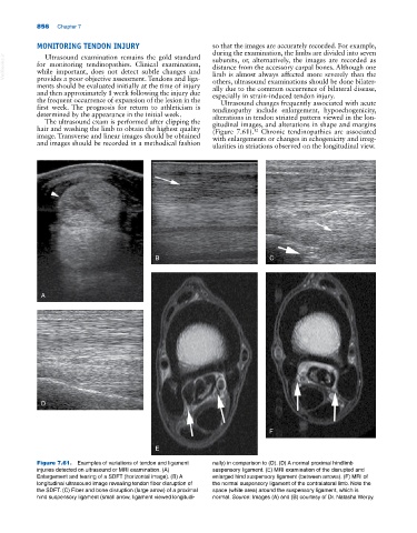
Figure 7.61. Examples of variations of tendon and ligament nally) in comparison to (D). (D) A normal proximal hindlimb
injuries detected on ultrasound or MRI examination. (A) suspensory ligament. (E) MRI examination of the disrupted and
Enlargement and tearing of a SDFT (horizontal image). (B) A enlarged hind suspensory ligament (between arrows). (F) MRI of
longitudinal ultrasound image revealing tendon fiber disruption of the normal suspensory ligament of the contralateral limb. Note the
the SDFT. (C) Fiber and bone disruption (large arrow) of a proximal space (white area) around the suspensory ligament, which is
hind suspensory ligament (small arrow, ligament viewed longitudi- normal. Source: Images (A) and (B) courtesy of Dr. Natasha Werpy.