
Page 892 - Adams and Stashak's Lameness in Horses, 7th Edition
P. 892
858 Chapter 7
VetBooks.ir
A B
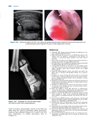
Figure 7.63. Ultrasound image of the DDFT with a linear tear located within the tendon sheath at the level of the fetlock (A) and the
corresponding tenoscopic image (B). The arrow in both images points to the tear.
References
1. Alexander RM. Energy‐saving mechanisms in walking and run
ning. J Exp Biol 1991;160:55–69.
2. Banes AJ, Tsuzaki M, Yamamoto J, et al. Mechanoreception at the
cellular level: the detection, interpretation, and diversity of
responses to mechanical signals. Biochem Cell Biol 1995;73:
349–365.
3. Barber SM. Arthrodesis of the distal intertarsal and tarsometatar
sal joints in the horse. Vet Surg 1984;13:227–236.
4. Birch HL, Rutter GA, Goodship AE. Oxidative energy metabolism
in equine tendon cells. Res Vet Sci 1997;62:93–97.
5. Cherdchutham W, Becker C, Smith RK, et al. Age‐related changes
and effect of exercise on the molecular composition of immature
equine superficial digital flexor tendons. Equine Vet J Suppl
1999;31:86–94.
6. Clegg PD. Musculoskeletal disease and injury, now and in the
future. Part 1: fractures and fatalities. Equine Vet J 2011;43:
643–649.
7. Dahlgren LA, van der Meulen MC, Bertram JE, et al. Insulin‐like
growth factor‐I improves cellular and molecular aspects of heal
ing in a collagenase‐induced model of flexor tendinitis. J Orthop
Res 2002;20:910–919.
8. Dahlgren LA, Mohammed HO, Nixon AJ. Temporal expression of
growth factors and matrix molecules in healing tendon lesions.
J Orthop Res 2005;23:84–92.
9. Dahlgren LA, Mohammed HO, Nixon AJ. Expression of insulin‐
like growth factor binding proteins in healing tendon lesions.
J Orthop Res 2006;24:183–92.
10. Dakin SG, Dudhia J, Smith RK. Resolving an inflammatory
concept: the importance of inflammation and resolution in tendi
nopathy. Vet Immunol Immunopathol 2014;158:121–127.
11. Dalton S, Cawston TE, Riley GP et al. Human shoulder tendon
biopsy samples in organ culture produce procollagenase and
tissue inhibitor of metalloproteinases. Ann Rheum Dis 1995;54:
571–577.
12. Davis CS, Smith RKW. Diagnosis and management of tendon and
ligament disorders. In Equine Surgery. Stick JA, Auer, JA, eds. W.B.
Saunders Co., Philadelphia, 2006;1086–1110.
Figure 7.64. Radiograph of a complete fetlock luxation 13. Dowling BA, Dart AJ. Mechanical and functional properties
demonstrating failure of the collateral ligaments. of the equine superficial digital flexor tendon. Vet J 2005;170:
184–192.
14. Dowling BA, Dart AJ, Hodgson DR, et al. Superficial digital flexor
tendonitis in the horse. Equine Vet J 2000;32:369–378.
result of trauma, characteristic stances of the limb can 15. Fortier LA, Smith RK. Regenerative medicine for tendinous and
often help the clinician determine which structure(s) ligamentous injuries of sport horses. Vet Clin North Am Equine
Pract 2008;24:191–201.
has(have) been affected (Figures 12.15 and 12.16). For 16. Genovese RL, Rantanen NW, Hauser ML, et al. Diagnostic ultra
more information on tendon lacerations, refer to sonography of equine limbs. Vet Clin North Am Equine Pract
Chapter 12. 1986;2:145–226.