
Page 883 - Adams and Stashak's Lameness in Horses, 7th Edition
P. 883
Principles of Musculoskeletal Disease 849
TENDON AND LIGAMENT INJURIES AND DISEASE
VetBooks.ir laurIe r. GoodrICh
ANATOMY glycosaminoglycan in the extracellular matrix. Larger
fibrils are stronger than smaller fibrils. 32
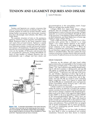
Tendons and ligaments are complex structures that Collagen fibrils have many triple helical collagen
exist in a hierarchical structure of subunits (Figure 7.52). molecules that are arranged such that a characteristic
Grossly, tendons are made up of many fascicles, which, banding pattern is seen on electron microscopy. Collagen
during further macroscopic and microscopic examina molecules are secreted extracellularly through the pores
tion, have decreasingly sized subunits, then fibers, and of the tenocytes. Cross‐linking of these molecules results
finally fibrils. in fibril formation, and these fibrils fuse as horses age,
The anatomic structures of note are the paratenon, leading to larger collagen fibrils. 17,21,32
epitenon, endotenon, tendon fascicles, and tendon fiber/ Under a light microscope, collagen fibers have a char
fibrils. Paratenon refers to the loose connective tissue acteristic wavy pattern that is referred to as crimp
and vessels that surround tendons. Epitenon is outside (Figure 7.53). Crimp imparts elasticity to the tendon.
of tendon fascicles and is continuous with the endote A decrease in crimp occurs with aging along with a
non. Endotenon contains vascular and neural structures greater reduction in the central fibers. 27,32,43 When the
and separate cell populations. Furthermore, the endote tendon is stretched, the central fibers straighten primar
non may be a source for pluripotential cells. Tendon fas ily, and therefore a greater load is placed on these fibers
cicles are the bundles of fibrocytes and tenocytes that relative to peripheral fibers. This may explain the more
are surrounded by endotenon. Tendon fibers/fibrils are common occurrence of tendon lesions observed centrally
made of long bundles of collagen filaments that are (core lesions). Peripheral lesions are less clearly explained.
predominately type I collagen and have elastin and
Cellular Components
Tropocollagen
Tenocytes are the primary cell types found within
Microfibril equine tendon; they are responsible for the formation and
maintenance of tendon tissue. Three types are described
32
(Figure 7.54). Type I cells have thin, spindle‐shaped nuclei,
Subfibril type II cells have rounded or oblong nuclei, and type III
cells appear as cartilage‐like cells with round nuclei and
visible nucleoli. Proportions vary with age of the horse,
32
site, and whether a ligament or tendon. Type I cells are
more frequently found in older horses, type II cells can be
Fibril found in higher numbers in young horses and in liga
Crimp
ments, and type III cells are found in areas sustaining
32
Endotenon higher compressive loads. It is logical to assume that
type II and III cells are metabolically active and maintain
tendon extracellular matrix; however, type I cells most
Paratenon Fasicle
likely do this to some extent as well.
Cellular numbers vary depending on age and location
within the tendon. As maturity is reached, the numbers
remain constant; however, areas of tendon become acel
lular and have changes consistent with chondroid meta
Tendon plasia such as the center of the superficial digital flexor
tendon (SDFT) or the deep digital flexor tendon (DDFT)
in the metacarpophalangeal region. These areas are
32
often surrounded by type II cells. Other cells found in
tendon include synovial‐like cells of the epitenon within
the tendon sheaths and fibroblasts of the paratenon, epi
tenon, and endotenon. These probably have an integral
role in tendon maintenance due to certain growth fac
Figure 7.52. A schematic representation of the tendon hierarchy. tors such as transforming growth factor‐β (TGF‐β)
8,18
The level of tendon fascicles can be viewed grossly, fibers may be found in these areas.
seen microscopically, and individual collagen fibrils (tropocollagen) Tenocyte regulation has not been fully elucidated,
may be viewed via electron microscopy. Source: Modified from Davis but most likely it relies on mechanical and cytokine
and Smith. Reproduced with permission of Elsevier. stimulation. Tenocytes have been shown to respond
12