
Page 878 - Adams and Stashak's Lameness in Horses, 7th Edition
P. 878
844 Chapter 7
VetBooks.ir
A B
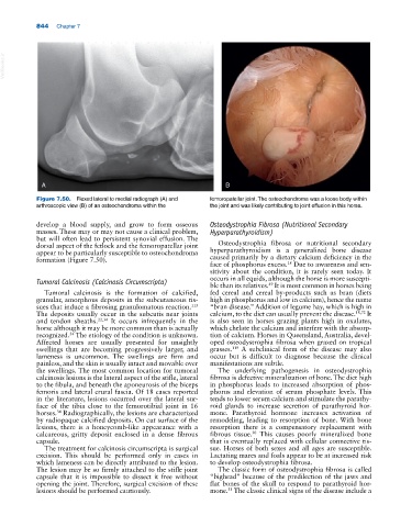
Figure 7.50. Flexed lateral to medial radiograph (A) and femoropatellar joint. The osteochondroma was a loose body within
arthroscopic view (B) of an osteochondroma within the the joint and was likely contributing to joint effusion in this horse.
develop a blood supply, and grow to form osseous Osteodystrophia Fibrosa (Nutritional Secondary
masses. These may or may not cause a clinical problem, Hyperparathyroidism)
but will often lead to persistent synovial effusion. The
dorsal aspect of the fetlock and the femoropatellar joint Osteodystrophia fibrosa or nutritional secondary
appear to be particularly susceptible to osteochondroma hyperparathyroidism is a generalized bone disease
formation (Figure 7.50). caused primarily by a dietary calcium deficiency in the
face of phosphorus excess. Due to awareness and sen
18
sitivity about the condition, it is rarely seen today. It
occurs in all equids, although the horse is more suscepti
Tumoral Calcinosis (Calcinosis Circumscripta)
49
ble than its relatives. It is most common in horses being
Tumoral calcinosis is the formation of calcified, fed cereal and cereal by‐products such as bran (diets
granular, amorphous deposits in the subcutaneous tis high in phosphorus and low in calcium), hence the name
sues that induce a fibrosing granulomatous reaction. “bran disease.” Addition of legume hay, which is high in
113
The deposits usually occur in the subcutis near joints calcium, to the diet can usually prevent the disease. 18,72 It
and tendon sheaths. 22,34 It occurs infrequently in the is also seen in horses grazing plants high in oxalates,
horse although it may be more common than is actually which chelate the calcium and interfere with the absorp
recognized. The etiology of the condition is unknown. tion of calcium. Horses in Queensland, Australia, devel
34
Affected horses are usually presented for unsightly oped osteodystrophia fibrosa when grazed on tropical
swellings that are becoming progressively larger, and grasses. A subclinical form of the disease may also
119
lameness is uncommon. The swellings are firm and occur but is difficult to diagnose because the clinical
painless, and the skin is usually intact and movable over manifestations are subtle.
the swellings. The most common location for tumoral The underlying pathogenesis in osteodystrophia
calcinosis lesions is the lateral aspect of the stifle, lateral fibrosa is defective mineralization of bone. The diet high
to the fibula, and beneath the aponeurosis of the biceps in phosphorus leads to increased absorption of phos
femoris and lateral crural fascia. Of 18 cases reported phorus and elevation of serum phosphate levels. This
in the literature, lesions occurred over the lateral sur tends to lower serum calcium and stimulate the parathy
face of the tibia close to the femorotibial joint in 16 roid glands to increase secretion of parathyroid hor
34
horses. Radiographically, the lesions are characterized mone. Parathyroid hormone increases activation of
by radiopaque calcified deposits. On cut surface of the remodeling, leading to resorption of bone. With bone
lesions, there is a honeycomb‐like appearance with a resorption there is a compensatory replacement with
calcareous, gritty deposit enclosed in a dense fibrous fibrous tissue. This causes poorly mineralized bone
30
capsule. that is eventually replaced with cellular connective tis
The treatment for calcinosis circumscripta is surgical sue. Horses of both sexes and all ages are susceptible.
excision. This should be performed only in cases in Lactating mares and foals appear to be at increased risk
which lameness can be directly attributed to the lesion. to develop osteodystrophia fibrosa.
The lesion may be so firmly attached to the stifle joint The classic form of osteodystrophia fibrosa is called
capsule that it is impossible to dissect it free without “bighead” because of the predilection of the jaws and
opening the joint. Therefore, surgical excision of these flat bones of the skull to respond to parathyroid hor
lesions should be performed cautiously. mone. The classic clinical signs of the disease include a
31