
Page 874 - Adams and Stashak's Lameness in Horses, 7th Edition
P. 874
840 Chapter 7
VetBooks.ir
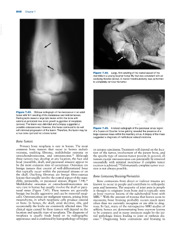
Figure 7.44. Large, firm swelling of the rostral aspect of the
mandible in a young Quarter horse filly that was consistent with an
ossifying fibroma (arrow). A rostral mandibulectomy was performed
to completely remove the tumor.
Figure 7.43. Oblique radiograph of the metatarsus in an adult
horse with firm swelling of the metatarsus and mild lameness.
Radiographs reveal a large lytic lesion within the bone with
extensive periosteal new bone growth suggestive of neoplasia
(arrows). The lesion was debrided and a biopsy suggested a
possible osteosarcoma. However, this horse continued to do well Figure 7.45. A lateral radiograph of the paranasal sinus region
with minimal progression of the lesion. Therefore, the lesion may be of a 3‐year‐old Quarter horse gelding revealed the presence of a
a true bone cyst and not a bone tumor. large osseous mass within the maxillary sinus. A biopsy of the mass
suggested a diagnosis of multilobular osteochondroma.
Bone Tumors
Primary bone neoplasia is rare in horses. The most
common bone tumors that occur in horses include or autopsy specimens. Treatment will depend on the loca
osteoma, ossifying fibroma, multilobular osteoma or tion of the tumor, involvement of the parent bone, and
36
osteochondrosarcoma, and osteosarcoma. Although the specific type of osseous tumor present. In general, all
these tumors may develop at any location, the face and tumors except osteosarcomas can potentially be removed
head (mandible, skull, and paranasal sinuses) appear to successfully with minimal recurrence if complete tumor
be the most common sites of occurrence. Osteomas are excision is achieved. Unfortunately complete tumor exci
36
benign tumors that consist of well‐differentiated bone sion is not always possible.
that typically occur within the paranasal sinuses or on
the skull. Ossifying fibromas are benign fibro‐osseous
lesions that usually involve the rostral aspect of the man Bone Contusion/Bruising/Periostitis
dible, premaxilla, or paranasal sinuses (Figure 7.44). Bone contusions from direct or indirect trauma are
36
Multilobular osteomas or osteochondrosarcomas are known to occur in people and contribute to orthopedic
very rare in horses but usually involve the skull or para pain and lameness. The majority of joint pain in people
nasal sinus (Figure 7.45). These tumors are generally is thought to originate from bone and is typically seen
benign but locally aggressive and can be removed surgi as bone marrow lesions of the subchondral bone with
cally. Osteosarcomas are malignant tumors, arising from MRI. With the amount of trauma that horses seem to
53
mesenchyme, in which neoplastic cells produce osteoid encounter, bone bruising probably occurs much more
or bone. In horses, the skull, axial skeleton, ribs, and often than we currently recognize or are able to diag
occasionally the limbs are commonly affected sites. The nose. In fact, many of the retrospective MRI studies of
clinical signs caused by these tumors will depend on the athletic horses are demonstrating bone marrow lesions
location and specific type of neoplasia. The diagnosis of to be common and in many instances might be the ini
neoplasia is usually made based on its radiographic tial pathologic lesion leading to joint or enthesis dis
appearance and is confirmed by histopathology of biopsy ease. Diagnosing bone contusions and bruising in
53