
Page 870 - Adams and Stashak's Lameness in Horses, 7th Edition
P. 870
836 Chapter 7
Osteomyelitis
Osteomyelitis is a more extensive inflammation of
VetBooks.ir the bone than osteitis that begins within or extends into
the medullary cavity. Osteomyelitis in horses can be
divided into three categories based on the origin of the
infection: hematogenous, traumatic, or iatrogenic.
7
Osteomyelitis from hematogenous origin occurs pri
marily in neonates and only rarely in adults. Traumatic
osteomyelitis can occur in any age horse and is usually
due to penetrating wounds or open fractures. Iatrogenic
causes of osteomyelitis include surgery such as internal
fixation of fractures or intra‐articular injections of
medications.
hematogenous InfeCtIon
The localization of hematogenous osteomyelitis in the
metaphyseal region of neonates can be explained by
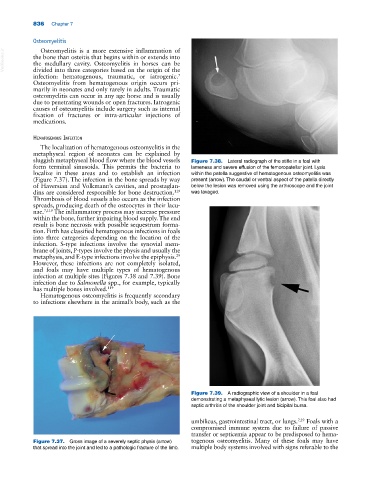
sluggish metaphyseal blood flow where the blood vessels Figure 7.38. Lateral radiograph of the stifle in a foal with
form terminal sinusoids. This permits the bacteria to lameness and severe effusion of the femoropatellar joint. Lysis
localize in these areas and to establish an infection within the patella suggestive of hematogenous osteomyelitis was
(Figure 7.37). The infection in the bone spreads by way present (arrow). The caudal or ventral aspect of the patella directly
of Haversian and Volkmann’s cavities, and prostaglan below the lesion was removed using the arthroscope and the joint
dins are considered responsible for bone destruction. was lavaged.
119
Thrombosis of blood vessels also occurs as the infection
spreads, producing death of the osteocytes in their lacu
nae. 7,119 The inflammatory process may increase pressure
within the bone, further impairing blood supply. The end
result is bone necrosis with possible sequestrum forma
tion. Firth has classified hematogenous infections in foals
into three categories depending on the location of the
infection. S‐type infections involve the synovial mem
brane of joints, P‐types involve the physis and usually the
metaphysis, and E‐type infections involve the epiphysis.
29
However, these infections are not completely isolated,
and foals may have multiple types of hematogenous
infection at multiple sites (Figures 7.38 and 7.39). Bone
infection due to Salmonella spp., for example, typically
has multiple bones involved. 119
Hematogenous osteomyelitis is frequently secondary
to infections elsewhere in the animal’s body, such as the
Figure 7.39. A radiographic view of a shoulder in a foal
demonstrating a metaphyseal lytic lesion (arrow). This foal also had
septic arthritis of the shoulder joint and bicipital bursa.
umbilicus, gastrointestinal tract, or lungs. 7,29 Foals with a
compromised immune system due to failure of passive
transfer or septicemia appear to be predisposed to hema
Figure 7.37. Gross image of a severely septic physis (arrow) togenous osteomyelitis. Many of these foals may have
that spread into the joint and led to a pathologic fracture of the limb. multiple body systems involved with signs referable to the