
Page 869 - Adams and Stashak's Lameness in Horses, 7th Edition
P. 869
Principles of Musculoskeletal Disease 835
are quite different, and it is important to make a distinc Sequestration without skin penetration does occur in
tion between the two categories of bone infection. 7 horses although it is rare. In cases in which there is no
VetBooks.ir Infectious Osteitis hematogenously leading to sequestrum formation, fistu
break in the skin, the hematoma may become infected
lation, drainage, and a nonhealing wound. This appears
Osteitis commonly occurs in the extremities of the to occur most commonly with injuries to the splint bones,
horse (mostly metacarpal/metatarsal regions) because of but most have skin wounds that lead to secondary bacte
the sparse soft tissue coverage in this location. It is usu rial infection. Chronic persistent drainage from any
ally the result of infection from a nearby septic process wound in the horse suggests the presence of a bone
or from a break in the skin. 7,10 Osteitis is seen frequently sequestrum or foreign body. Drainage will rarely subside
when a horse is kicked without breaking the overlying or at least wound healing will be substantially prolonged
skin and may be similar to a bone bruise when no without surgical removal of the sequestrum. This is
sequestrum develops. If the skin is broken, exposing the because the pathogenic organisms reside within the
periosteum, the outer layers of cortical bone may even necrotic bone, which is avascular, thereby resisting the
tually die, whereas the deeper cortical layers of bone animal’s immune defenses.
survive due to the blood supply from endosteal vessels. The severity of lameness accompanying osteitis in
For example, lacerations with bone exposure of the dor horses is variable and inconsistent. In addition, the
sal aspect of the metacarpus/metatarsus frequently radiographic signs of osteitis will depend on the dura
develop osteitis and sequestration. 7,10 Bacteria that gain tion that has elapsed between the injury and time of
entrance to the bone lodge in the superficial layers of the examination. Initially there may be soft tissue swelling
bone, resulting in a thin layer of dead bone (bone seques with evidence of bone resorption seen radiographi
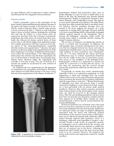
trum) within the wound (Figure 7.36). Although granu cally. At 7–14 days following the injury, periosteal
lation tissue may advance over the bone sequestrum, the proliferation may be evident. Sequestrum formation
rate of advancement is usually slow. Occasionally gran may also be visible at that time as osteoclastic resorp
ulation tissue advances under the sequestrum and tion occurs at the periphery of the damaged bone.
extrudes it from the wound. The rate of healing of a Radiographic evidence of a sequestrum is usually not
wound can usually be accelerated by early removal of visible for a minimum 2–3 weeks after the injury. At
10
the sequestrum. this time, the sequestrum and the sclerotic margin
By definition the two requirements for the formation around the sequestrum called the involucrum are usu
of a sequestrum are avascularity and infection. Therefore, ally visible. 7
most surgeons feel that blunt trauma to the bone cortex Occasionally an osteitis may resolve spontaneously,
does not cause sequestration in the absence of infection. 7,10 especially if there is no infectious component or if the
sequestrum is small and extruded from the wound
(which rarely occurs on its own). If bacteria and necrotic
bone are present, the wound will be exudative indefinitely
119
until the sequestrum is removed. Wound debridement
of unhealthy scar tissue and necrotic bone is usually
required for healing to occur. Removal of bone seques
tra is best performed with the animal under general
anesthesia. However, thin cortical sequestra associated
with avulsion injuries of the dorsal aspect of the cannon
bone can often be removed in the standing sedated
horse. After the surrounding granulation and scar tissue
have been excised, the area should be curetted until the
bone appears to be healthy. Most wounds are either
closed primarily or left to heal by second intention fol
lowing debridement.
Parenteral antibiotics are of limited value when used
alone to treat bone sequestra because of poor penetra
tion of the antibiotics into the necrotic bone. Antibiotics
are indicated if there are signs of cellulitis (phlegmon)
associated with the lesion and following surgical
debridement of the wound. Occasionally a mild seques
trum can be effectively treated with antibiotics admin
istered through regional limb perfusion, which is a
technique commonly used regardless if surgery is per
formed. Usually a wide variety of organisms (second
ary pathogens) can be cultured from the wound, and
occasionally these bacteria are resistant to antibiotics
that are of practical use in the horse. Culturing the
sequestrum itself usually gives the most accurate diag
nosis as to the causative bacteria. The prognosis for
Figure 7.36. A sequestrum on the lateral aspect of the fourth horses with osteitis and sequestrum removal is usually
metatarsal bone (arrow) secondary to trauma. excellent.