
Page 872 - Adams and Stashak's Lameness in Horses, 7th Edition
P. 872
838 Chapter 7
established where nutrients are available, proliferation
occurs within a polysaccharide slime, forming a biofilm‐
VetBooks.ir bacterial extracapsular exopolysaccharide that binds to
enclosed colony. This biofilm or bioslime is formed by
surfaces of the implants and help maintain infection by
protecting the bacteria from the host defenses. Because
of this, osteomyelitis may develop despite prophylactic
antibiotic coverage at the time of fracture repair espe
cially in open fractures. Highly resistant bacteria such as
methicillin‐resistant Staphylococcus aureus or Gram‐
negative enterics often cause these types of infections. 7
ClInICal sIgns of osteomyelItIs
Hematogenous osteomyelitis may be missed in its
early stages and often presents after the lameness has
become unresponsive to medical therapy. Frequently the
owner feels that the lameness was due to an injury such
as a sprain or being stepped on by the mare. There is
usually a very severe lameness with cellulitis similar to
that seen with fractures. Pain is usually elicited with
direct pressure and manipulation of the joint(s) adjacent
to the infection, and a fever is commonly seen in foals.
Clinical signs of traumatic or iatrogenic osteomyelitis
are similar and include lameness, soft tissue swelling,
and retarded wound healing over the implants, drain
age, and fistulation. Signs can be present as early as
7–10 days of injury or surgery or may be delayed for
3–4 weeks. Bloodwork usually demonstrates most con
sistently an increase in fibrinogen and occasionally
increased white blood cell count. Serum amyloid A is
often increased dramatically in these cases.
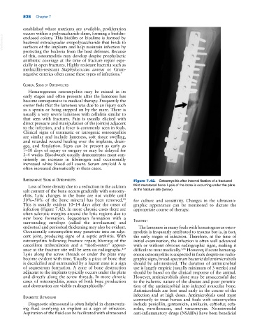
radIograPhIC sIgns of osteomyelItIs Figure 7.42. Osteomyelitis after internal fixation of a fractured
Loss of bone density due to a reduction in the calcium third metatarsal bone. Lysis of the bone is occurring under the plate
salt content of the bone occurs gradually with osteomy at the fracture site (arrow).
elitis. Lytic changes in the bone are not visible until
89
30%–50% of the bone mineral has been removed. for culture and sensitivity. Changes in the ultrasono
This is usually evident 10–14 days after the onset of graphic appearance can be monitored to dictate the
infection (Figure 7.42). In more chronic cases there are appropriate course of therapy.
often sclerotic margins around the lytic regions due to
new bone formation. Sequestrum formation with a
surrounding envelope (called the involucrum) and treatment
endosteal and periosteal thickening may also be evident. The lameness in many foals with hematogenous osteo
Occasionally osteomyelitis may penetrate into an adja myelitis is frequently attributed to trauma but is, in fact,
cent joint, producing signs of a septic arthritis. With the early stages of infection. Therefore, at the time of
osteomyelitis following fracture repair, blurring of the initial examination, the infection is often well advanced
cancellous trabeculation and a “moth‐eaten” appear with or without obvious radiographic signs, making it
ance at the fracture site will be seen on radiographs. difficult to treat medically. 7,96 However, if acute hematog
119
Lysis along the screw threads or under the plate may enous osteomyelitis is suspected in foals despite no radio
become evident with time. Usually a piece of bone that graphic signs, broad‐spectrum bactericidal antimicrobials
is decalcified and surrounded by a lucent zone is a sign should be administered. The duration of antimicrobial
of sequestrum formation. A zone of bone destruction use is largely empiric (usually minimum of 3 weeks) and
adjacent to the implants typically occurs under the plate should be based on the clinical response of the animal.
and directly along the screw threads. In more chronic However, antimicrobials alone may be unsuccessful due
cases of osteomyelitis, zones of both bone production to the ischemic nature of the disease and poor penetra
and destruction are visible radiographically. 7 tion of the antimicrobial into infected avascular bone.
Antimicrobials are best used early in the course of the
infection and at high doses. Antimicrobials used most
dIagnostIC ultrasound
commonly to treat horses and foals with osteomyelitis
Diagnostic ultrasound is often helpful in characteriz include penicillin, gentamicin, amikacin, ceftiofur, cefa
ing fluid overlying an implant as a sign of infection. zolin, enrofloxacin, and vancomycin. Nonsteroidal
Aspiration of the fluid can be facilitated with ultrasound anti‐inflammatory drugs (NSAIDs) have been beneficial