
Page 871 - Adams and Stashak's Lameness in Horses, 7th Edition
P. 871
Principles of Musculoskeletal Disease 837
VetBooks.ir
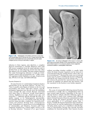
Figure 7.40. Radiographs of the fetlock joint from a mare that
had a penetrating injury to the fetlock joint 2 weeks previously. Note
the severe subchondral bone lysis (arrowhead) and joint space
collapse (arrow) in the joint secondary to sepsis. Figure 7.41. An oblique radiograph of the tarsus in a horse with
a puncture wound and infection of the calcaneal bursa. Lysis within
the proximal aspect of the calcaneus (black arrows) suggested
infection in these regions, and, therefore, a complete concurrent osteitis or osteomyelitis of the bone.
physical examination is essential. However, some foals
will recover completely from the initial infection only to
develop bone or joint infections several days later when without providing absolute stability is usually futile
they appear to be very healthy. The most common bacteria unless the dead avascular fragments can be removed or
that cause hematogenous infections in foals are Gram‐ reincorporated into the healing fracture where they can
negative enterics such as Escherichia coli. 7,29 Other causa be revascularized. Alternatively, some type of external
119
tive organisms include Staphylococcus spp., Streptococcus fixator may be used to stabilize the fracture without dis
spp., Rhodococcus spp., and Salmonella spp. rupting the soft tissues around the fracture site. 69,80,82 In
this manner, the potential for osteomyelitis is decreased
traumatIC osteomyelItIs because no implants are placed near the fracture, and
the vascularity to the fracture is not further impaired.
An open fracture or a penetrating wound may lead to
osteomyelitis in any age horse (Figures 7.40 and 7.41).
There is usually some degree of trauma to the skin and
surrounding soft tissues with these injuries, and the IatrogenIC osteomyelItIs
pathogenic organisms may directly enter the medullary The cause of osteomyelitis following internal fixation
cavity through the open wound. Bacteria associated of fractures is usually due to contamination from an
with these types of infections include Gram‐negative open wound (open fracture). Regardless of the type of
enterics, Staphylococcus spp., Streptococcus spp., and internal fixation utilized, infection following repair of
anaerobes. 7,117 Infection spreads through the bone in a open fractures is much more common than following
similar manner as for hematogenous osteomyelitis. repair of closed fractures. However, contamination of the
Occasionally there is no overt break in the skin, but the fracture during the surgical procedure can and does
necrotic tissue provides a medium for bacterial prolif occur, particularly if it is prolonged (greater than 3
7
eration and infection develops from a hematogenous hours). Usually the fracture hematoma and avascularity
route. Avascularity is a major factor in the pathogenesis at the fracture site and the implantation of foreign mate
of osteomyelitis, and therefore, fractures with bone rial (pins, plates, screws, etc.) contribute to the develop
fragments isolated from a blood supply will be at risk ment of osteomyelitis, because they provide favorable
to develop infection. Treatment of an open fracture conditions for bacterial growth. Once bacteria become
7