
Page 875 - Adams and Stashak's Lameness in Horses, 7th Edition
P. 875
Principles of Musculoskeletal Disease 841
VetBooks.ir
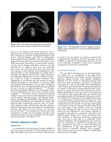
Figure 7.46. MR image of the distal phalanx demonstrating
marrow edema (arrow). Source: Courtesy of Dr. Kurt Selberg. Figure 7.47. The distal aspect of the third metacarpal condyles
typically shows subchondral bone bruising in racing Thoroughbred
horses (arrow).
horses is very difficult and mostly subjective. This is
especially true for bone pain originating in joints. Most
bone contusions or bruises are diagnosed based on the
history, clinical findings, and lack of radiographic in horses is an uncommon or at least an uncommonly
abnormalities. However, MRI is now used routinely to recognized clinical problem. The term osteopenia is used
diagnose bruising and edema within bone (Figure 7.46). if BMD is reduced, but spontaneous fractures do not
With some bone contusions of cortical bone, evidence occur. Clinically, osteopenia is much more commonly
of pain can be elicited with direct pressure over the recognized in horses than osteoporosis.
affected site. In addition, bone pain can sometimes
cause more severe lameness than most soft tissue inju
ries, and pain is often elicited with manipulation of the Generalized Osteoporosis
affected bone or joint. Radiographs are useful only to The generalized osteoporosis seen in postmenopau
document the absence of fractures or other abnormali sal women has no counterpart in the aged horse.
ties within the affected bone or joint. Nuclear scintigra However, older mares may be more prone to long bone
phy would be useful to document abnormal bone fractures during recovery from general anesthesia.
metabolism of the affected site, but cannot be used to Whether this may be due to declining estrogen levels
definitively diagnose a bone bruise or contusion. MRI associated with reproductive senescence is unknown.
and computed tomography (CT) are used in people to Osteoporosis is seen occasionally with undernutrition
help diagnose bone damage/contusion and are available rather than actual deficiencies of calcium, phosphorus,
for use in horses at referral hospitals. 66,73,108 Positron or vitamin D. However, in most affected horses, osteo
emission tomography (PET) has also been used to iden porosis is usually associated with a diet low in calcium,
tify potential lesions, but more work is needed on this high in phosphorus, or low in vitamin D. Osteoporosis
modality. 109,110 Treatment of bone contusions would be is associated with copper deficiency and chronic lead
similar to any type of acute musculoskeletal trauma and poisoning in lambs, but this has not been seen in foals.
9
would include reduced activity, cold therapy, hydro A condition first recognized in Thoroughbred foals may
therapy, bandaging, and NSAIDs. Suspected bone con be a manifestation of generalized osteoporosis. The
26
tusions/bruising within joints could also be treated with condition is characterized by fractures of the proximal
intra‐articular medications; however, they are some sesamoid bones and typically occurs when foals gallop
what limited in their effectiveness. Systemic bisphos to exhaustion trying to keep up with their dams. Foals
phonate therapy has been advocated for treating bone seem more prone to the condition if they are confined
bruising and edema; however efficacy has not been after birth. During this time of relative inactivity, the
proven in horses. The prognosis of horses with bone bones are not subjected to the stresses required to
50
contusions is usually very good unless damage to the strengthen them and are a potential weak link in the
subchondral bone of a joint contributes to joint pathol skeletal system (Figure 7.21). Some underlying meta
ogy and secondary OA (Figure 7.47). bolic problem or deficiency producing osteoporosis
may exist, but at this time it is undefined. Other
lameness problems and unexplained fractures in horses
SYSTEMIC DISEASES OF BONE may be attributed to generalized osteoporosis, but
Osteoporosis their pathogenesis and cause are often unexplainable.
The combination of age and pregnancy have also
In osteoporosis, the bone mineral density (BMD) of been shown to negatively influence bone strength,
37
the bone matrix is reduced. The bone becomes porous, with increased age and parity leading to significantly
light, and fragile and is prone to fracture. Osteoporosis decreased bone strength.