
Page 876 - Adams and Stashak's Lameness in Horses, 7th Edition
P. 876
842 Chapter 7
Further experiments have shown that treatment of
Localized Osteopenia (Disuse Osteopenia) osteopenia with 25‐hydroxycholecalciferol has been
This condition is fairly common, especially in horses
VetBooks.ir following rigid external immobilization of their limbs beneficial. Fortunately, localized osteopenia following
25
immobilization rarely causes any problems. If the external
(casting). Disuse osteopenia may also occur in horses
with severe or chronic lameness (Figure 7.48) or neu
ropathies such as radial nerve paralysis where weight‐
bearing is reduced. With decreased weight‐bearing, there
is increased resorption of bone and decreased bone for
mation. Immobilization of the thoracic limb of a pony in
a cast for 6 weeks caused a significant decrease in weight
and specific gravity of the third metacarpal bone.
25
Histologically, osteopenia was caused by atrophy of
osteoblasts with failure of bone apposition. 25,119
However, external immobilization of the distal limb in
horses has also been shown to have only a minor effect
on articular cartilage with very little clinical signifi
cance. If horses are brought back into work too
93
quickly, distal sesamoidean ligaments can tear from the
distal aspect of the proximal sesamoid bones, leading to
significant lameness (Figure 7.21). Osteopenia is usually
more severe in young animals due to the inherent rapid
bone turnover compared with the adult horse. Bone cor
tices become thinner and more lytic. Although this is
thought to rarely contribute to a clinical problem and is
easily reversed when the external immobilization device
is removed and the animal commences normal weight‐
bearing, recent evidence suggests that the pathologic
changes can be significant.
Localized osteopenia is essentially a radiographic
diagnosis characterized by lack of cortical density and a
more lucent appearance to the bones compared with
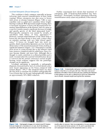
normal (Figure 7.49). The sesamoid bones are often Figure 7.48. A lateromedial radiograph showing a comminuted
119
the first bone to manifest the problem radiographically. fracture of the femur in a 23‐year‐old mare. This mare had severe
Loss of bone that can be seen radiographically indicates lameness in the stifle of that limb since she was a yearling and fractured
an approximately 30% BMD reduction. it while getting up in her stall. It is likely that the bone was osteoporotic
due to chronic, reduced weight‐bearing from the lameness.
A B C
Figure 7.49. Radiographic images of a comminuted P2 fracture months after pin removal. Note the progression of disuse osteoporo-
that was treated with a transfixation pin cast. (A) At the initial pin sis of the sesamoid bones (arrows in A, B, and C) and distal limb
placement. (B) When the pins were removed 3 months later. (C) Five with subsequent fracturing of the sesamoid bones (C, arrow).