
Page 879 - Adams and Stashak's Lameness in Horses, 7th Edition
P. 879
Principles of Musculoskeletal Disease 845
symmetrical enlargement of the mandible and facial associated with intra‐abdominal disorders without pul
bones. There is loss of lamina dura around the teeth due monary involvement. Hypertrophic osteopathy has been
VetBooks.ir teeth may eventually loosen. This can be identified radi ovarian primordial germ cells) that had abdominal
reported in a mare with a dysgerminoma (a neoplasm of
to osteoclastic resorption of alveolar margins, and the
metastases but was free of thoracic lesions.
ographically and is one of the first signs of the disease.
67
Swelling initially begins just above the facial crest and in The two most common mechanisms for the develop
the mandible producing a reduction in intermandibular ment of the disease are classified as neurogenic and
space. There may also be enough swelling of the palate, humoral. The more popular neurogenic theory is based
maxillae, and incisor bones to produce dyspnea. on the fact that an apparent stimulation of the vagus
The subclinical form of nutritional secondary hyper nerve produces an alteration of the vasculature and peri
parathyroidism may cause a nebulous shifting limb lame osteum of bones by way of an unknown efferent pathway.
ness in horses that is difficult to localize with standard Support for this theory is based on the fact that in dogs
perineural anesthesia. The condition may also contrib and man hypertrophic osteopathy lesions may regress fol
119
ute to unexplained fractures especially in young horses. In lowing vagotomy. A humoral mechanism may also exist,
such cases a dietary history is important in the diagnosis. since hypertrophic osteopathy has been shown to occur in
Blood calcium and phosphorus levels are usually normal, people when urinary excretion of estrogen is increased.
and the diagnosis is usually based on analysis of the diet High levels of circulating estrogen have been reported in
and response to treatment with calcium supplementation. a mare with hypertrophic osteopathy, although the exact
relationship between estrogen levels and hypertrophic
Hypertrophic Osteopathy (Hypertrophic Pulmonary osteopathy is purely speculative. 67
Osteoarthropathy) The clinical signs of hypertrophic osteopathy are related
to periosteal hyperostoses. There is symmetric enlargement
Hypertrophic osteopathy is a progressive bilaterally of the long bones of the limb with all bones in the limb
symmetrical proliferation of subperiosteal bone and affected. There is pain and edema of soft tissues, and the
fibrous connective tissue on the appendicular and axial horse may have a stiff gait and be reluctant to move. The
skeleton and facial bones. It is a relatively rare disease joint surfaces are rarely involved; however, there may be
119
in the horse. The pathogenesis of hypertrophic osteopa decreased motion in affected joints as well as pain on
thy is still unclear at present. Classically the disease is manipulation. There may be signs referable to pulmonary
associated with a space‐occupying pulmonary lesion abnormalities such as coughing and nasal discharge.
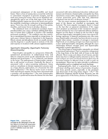
such as a neoplasm or chronic suppurative process such Radiographically, there is a generalized increase in
as a large abscess, tuberculosis, or a fractured rib with soft tissue swelling and evidence of periostitis. Irregular
67
pleural adhesions. The disease rarely occurs in horses new bone growth occurs, especially at the proximal
with such thoracic lesions, but has been associated with and distal ends of the long bones (Figure 7.51). The
119
32
a granular cell myoblastoma. The term hypertrophic differential diagnosis should include fluorosis, but the
osteopathy is preferred because the disease has also been gross appearance of the bones, absence of dental lesions,
Figure 7.51. Lateral (A) and
dorsopalmar (B) radiographs of the left
front fetlock of a horse with hypertrophic
osteopathy showing irregular new bone
growth. A definitive cause was never A B
determined.