
Page 643 - Equine Clinical Medicine, Surgery and Reproduction, 2nd Edition
P. 643
618 CHAPTER 3
VetBooks.ir 3.45 3.46
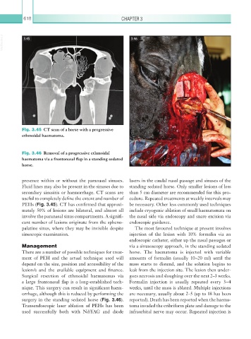
Fig. 3.45 CT scan of a horse with a progressive
ethmoidal haematoma.
Fig. 3.46 Removal of a progressive ethmoidal
haematoma via a frontonasal flap in a standing sedated
horse.
presence within or without the paranasal sinuses. lasers in the caudal nasal passage and sinuses of the
Fluid lines may also be present in the sinuses due to standing sedated horse. Only smaller lesions of less
secondary sinusitis or haemorrhage. CT scans are than 5 cm diameter are recommended for this pro-
useful to completely define the extent and number of cedure. Repeated treatments at weekly intervals may
PEHs (Fig. 3.45). CT has confirmed that approxi- be necessary. Other less commonly used techniques
mately 50% of lesions are bilateral, and almost all include cryogenic ablation of small haematomata on
involve the paranasal sinus compartments. A signifi- the nasal side via endoscopy and snare excision via
cant number of lesions originate from the spheno- endoscopic guidance.
palatine sinus, where they may be invisible despite The most favoured technique at present involves
sinuscopic examination. injection of the lesion with 10% formalin via an
endoscopic catheter, either up the nasal passages or
Management via a sinusoscopy approach, in the standing sedated
There are a number of possible techniques for treat- horse. The haematoma is injected with variable
ment of PEH and the actual technique used will amounts of formalin (usually 10–20 ml) until the
depend on the size, position and accessibility of the mass starts to distend, and the solution begins to
lesion/s and the available equipment and finance. leak from the injection site. The lesion then under-
Surgical resection of ethmoidal haematomas via goes necrosis and sloughing over the next 2–3 weeks.
a large frontonasal flap is a long-established tech- Formalin injection is usually repeated every 3–4
nique. This surgery can result in significant haem- weeks, until the mass is ablated. Multiple injections
orrhage, although this is reduced by performing the are necessary, usually about 2–5 (up to 18 has been
surgery in the standing sedated horse (Fig. 3.46). reported). Death has been reported when the haema-
Transendoscopic laser ablation of PEHs has been toma invaded the cribriform plate and damage to the
used successfully both with Nd:YAG and diode infraorbital nerve may occur. Repeated injection is