
Page 645 - Equine Clinical Medicine, Surgery and Reproduction, 2nd Edition
P. 645
620 CHAPTER 3
VetBooks.ir 3.48 extensive before they are diagnosed, making suc-
cessful treatment unlikely, the chance of recurrence
high and the long-term prognosis poor. Benign
lesions that are well circumscribed can be success-
fully treated by conventional surgical excision, laser
ablation or even intralesional formalin injections.
PARANASAL SINUS CYSTS
Definition/overview
Swelling rostral to the rostral edge of the facial crest
is frequently caused by dental disease. By contrast,
swelling caudal to the rostral edge of the facial crest
is rarely caused by dental disease and is almost invari-
ably caused by an expansile mass within the parana-
sal sinuses. Paranasal sinus cysts are one of the most
frequent expansile lesions of this area. The cause of
the condition is unclear and the precise pathology is
debated.
Aetiology/pathophysiology
The aetiology and pathophysiology are unknown.
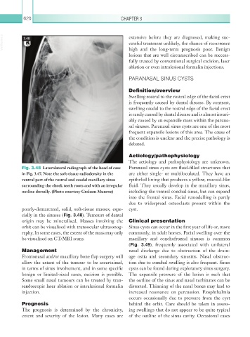
Fig. 3.48 Laterolateral radiograph of the head of case Paranasal sinus cysts are fluid-filled structures that
in Fig. 3.47. Note the soft-tissue radiodensity in the are either single- or multiloculated. They have an
ventral part of the rostral and caudal maxillary sinus epithelial lining that produces a yellow, mucoid-like
surrounding the cheek teeth roots and with an irregular fluid. They usually develop in the maxillary sinus,
outline dorsally. (Photo courtesy Graham Munroe) including the ventral conchal sinus, but can expand
into the frontal sinus. Facial remodelling is partly
due to widespread osteoclasts present within the
poorly-demarcated, solid, soft-tissue masses, espe- cyst.
cially in the sinuses (Fig. 3.48). Tumours of dental
origin may be mineralised. Masses involving the Clinical presentation
orbit can be visualised with transocular ultrasonog- Sinus cysts can occur in the first year of life or, more
raphy. In some cases, the extent of the mass may only commonly, in adult horses. Facial swelling over the
be visualised on CT/MRI scans. maxillary and conchofrontal sinuses is common
(Fig. 3.49), frequently associated with unilateral
Management nasal discharge due to obstruction of the drain-
Frontonasal and/or maxillary bone flap surgery will age ostia and secondary sinusitis. Nasal obstruc-
allow the extent of the tumour to be ascertained, tion due to conchal swelling is also frequent. Sinus
in terms of sinus involvement, and in some specific cysts can be found during exploratory sinus surgery.
benign or limited-sized cases, excision is possible. The expansile pressure of the lesion is such that
Some small nasal tumours can be treated by tran- the outline of the sinus and nasal turbinates can be
sendoscopic laser ablation or intralesional formalin distorted. Thinning of the nasal bones may lead to
injection. increased resonance on percussion. Exophthalmia
occurs occasionally due to pressure from the cyst
Prognosis behind the orbit. Care should be taken in assess-
The prognosis is determined by the chronicity, ing swellings that do not appear to be quite typical
extent and severity of the lesion. Many cases are of the outline of the sinus cavity. Occasional cases