
Page 644 - Equine Clinical Medicine, Surgery and Reproduction, 2nd Edition
P. 644
Respir atory system: 3.2 Surgical conditions of the respir atory tr act 619
VetBooks.ir more complicated in horses when the mass is within tumours derived from tooth-forming elements are
rare and usually occur in young animals.
the paranasal sinuses. Intermittent injection is fea-
sible but repeated trephination becomes traumatic to
the horse. Injection within the sinuses can result in Clinical presentation
significant chemical sinusitis, although this usually The clinical signs vary with the location and extent
resolves with medical management. of the mass. Early localised cases may be identified
incidentally or be presented because of localised
Prognosis secondary infection of the mass. Drainage from the
The prognosis is guarded. Recurrence is common, paranasal sinuses may be affected, leading to a sec-
approximately 15–50% of cases within a 1–2 year- ondary sinusitis. Nasal discharge is often foul smell-
period, either due to re-growth, incomplete removal ing, bloody, purulent and usually unilateral unless
or new lesions. It has been suggested that the prog- the mass is so extensive as to invade the other side
nosis is better following CT scans, as hitherto unde- of the head. Expansion of the mass can lead to facial
tected lesions can be treated. Follow-up endoscopy swelling, airway obstruction and exophthalmos.
every 6 months is recommended to detect early Those tumours involving the mouth may present
recurrence Repeated injection with formalin or with halitosis and occasionally dysphagia. In rare
transendoscopic laser ablation is feasible. Many cases cases the mass may appear at the nares.
will be lost to follow-up due to client frustration at
repeated veterinary attention and expense, with lim- Differential diagnosis
ited prospect of a permanent cure. Other causes of secondary sinusitis, including max-
illary cysts, chronic primary sinus empyema, PEH
SINUS AND NASAL NEOPLASIA and nasal polyps, should be considered.
Definition/overview Diagnosis
Tumours of the nasal passages and paranasal sinuses A thorough clinical examination including evalu-
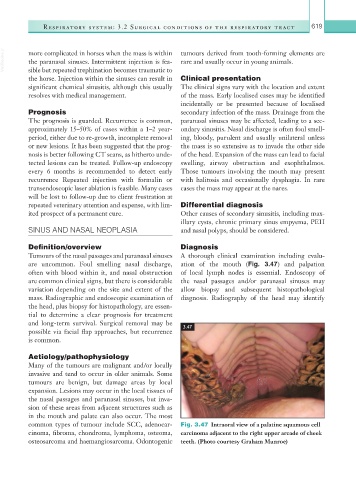
are uncommon. Foul smelling nasal discharge, ation of the mouth (Fig. 3.47) and palpation
often with blood within it, and nasal obstruction of local lymph nodes is essential. Endoscopy of
are common clinical signs, but there is considerable the nasal passages and/or paranasal sinuses may
variation depending on the site and extent of the allow biopsy and subsequent histopathological
mass. Radiographic and endoscopic examination of diagnosis. Radiography of the head may identify
the head, plus biopsy for histopathology, are essen-
tial to determine a clear prognosis for treatment
and long-term survival. Surgical removal may be
possible via facial flap approaches, but recurrence 3.47
is common.
Aetiology/pathophysiology
Many of the tumours are malignant and/or locally
invasive and tend to occur in older animals. Some
tumours are benign, but damage areas by local
expansion. Lesions may occur in the local tissues of
the nasal passages and paranasal sinuses, but inva-
sion of these areas from adjacent structures such as
in the mouth and palate can also occur. The most
common types of tumour include SCC, adenocar- Fig. 3.47 Intraoral view of a palatine squamous cell
cinoma, fibroma, chondroma, lymphoma, osteoma, carcinoma adjacent to the right upper arcade of cheek
osteosarcoma and haemangiosarcoma. Odontogenic teeth. (Photo courtesy Graham Munroe)