
Page 232 - Anatomy and Physiology of Farm Animals, 8th Edition
P. 232
Physiology of the Nervous System / 217
resistance to the flow of current. The largest between the cell membranes of adjacent
neurons that permit ionic exchange.
(as much as 20 μm in diameter), most heavily
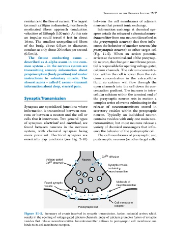
VetBooks.ir myelinated fibers approach conduction Information exchange at chemical syn
velocities of 250 mph (130 m/s). At this rate
transmitter from one neuron (described as
an impulse could travel 6 feet in about apses entails the release of a chemical neuro-
16 ms. The smallest unmyelinated fibers the presynaptic neuron) that then influ
of the body, about 0.5 μm in diameter, ences the behavior of another neuron (the
conduct at only about 20 inches per second postsynaptic neuron) or other target cell
(0.5 m/s). (Fig. 11‐5). When an action potential
The fastest conducting axons – arrives at the terminal end of the presynap
described as A‐alpha axons in one com- tic neuron, the change in membrane poten
mon system – in the nervous system are tial is responsible for opening voltage‐gated
transmitting sensory information about calcium channels. The calcium concentra
proprioception (body position) and motor tion within the cell is lower than the cal
instructions to voluntary muscle. The cium concentration in the extracellular
slowest axons – called C axons – transmit fluid, so calcium will flow through the
information about deep, visceral pain. open channels into the cell down its con
centration gradient. The increase in intra
cellular calcium within the terminal end of
Synaptic Transmission the presynaptic neuron sets in motion a
complex series of events culminating in the
Synapses are specialized junctions where release of neurotransmitters stored in
information is transmitted between neu secretory vesicles within the presynaptic
rons or between a neuron and the cell or neuron. Typically, an individual neuron
cells that it innervates. Two general types contains vesicles with only one main neu
of synapses, electrical and chemical, are rotransmitter, but most neurons release a
found between neurons in the nervous variety of chemical messengers that influ
system, with chemical synapses being ence the behavior of the postsynaptic cell.
more prevalent. Electrical synapses are The cell membranes of presynaptic and
essentially gap junctions (see Fig. 2‐10) postsynaptic neurons (or other target cells)
Ca 2+ diffusion
Voltage-gated Neuron
Ca 2+ channel
Synaptic vesicle
containing
neurotransmitter
Fused synaptic Molecule of
vesicle neurotransmitter
Cell membrane
Postsynaptic cell receptor
Figure 11-5. Summary of events involved in synaptic transmission. Action potential arrives which
results in the opening of voltage‐gated calcium channels. Entry of calcium promotes fusion of synaptic
vesicles that release neurotransmitter. Neurotransmitter diffuses to postsynaptic cell membrane and
binds to its cell membrane receptor.