
Page 237 - Anatomy and Physiology of Farm Animals, 8th Edition
P. 237
222 / Anatomy and Physiology of Farm Animals
One specific type of reflex arc requires
Reflexes Involving Skeletal Muscle only two neurons, with the sensory neuron
VetBooks.ir Contraction synapsing directly on the motor neuron;
with only one synapse between the input
A reflex is a stereotyped response to a given
stimulus which can operate without con and output limbs of the reflex, this mono-
scious/voluntary influence. To say that the synaptic reflex provides an extremely fast
response is “stereotyped” means that it is response. The only circumstance in which
the same each time the reflex is activated.
Reflexes exist to maintain a stable internal
environment (e.g., increases in heart rate CNS PNS
with increased oxygen depletion during Cell body in sensory ganglion
exercise), to provide fast postural correc
tions (e.g., contraction of a muscle that is
stretched due to an unexpected load), or to Receptor
protect the individual from harm (e.g., with
drawing a limb from a painful stimulus).
The vast majority of reflexes consist Interneuron Sensory neuron
of: (1) a sensory limb, which comprises a
receptor and sensory neuron; (2) central
integration via interneuron(s); and (3) a Target
motor limb, comprising a motor neuron
and the target of the reflex (Figs. 11‐8 and Motor neuron
11‐9). Reflexes may produce contraction of
skeletal muscle or, in the case of autonomic
reflexes, changes in the behavior of smooth
muscle, cardiac muscle, or glands. Figure 11-8. Schematic of a typical reflex arc.
(A)
(B)
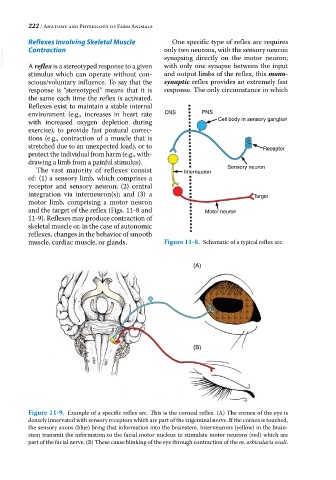
Figure 11-9. Example of a specific reflex arc. This is the corneal reflex. (A) The cornea of the eye is
densely innervated with sensory receptors which are part of the trigeminal nerve. If the cornea is touched,
the sensory axons (blue) bring that information into the brainstem. Interneurons (yellow) in the brain
stem transmit the information to the facial motor nucleus to stimulate motor neurons (red) which are
part of the facial nerve. (B) These cause blinking of the eye through contraction of the m. orbicularis oculi.